Liu Xiaoli, Lin Huichao, Song Jiaao, Zhang Taiyi, Wang Xiaoying, Huang Xiaowen, Zheng Chengyun
Department of Hematology, The Second Hospital, Cheeloo College of Medicine, Shandong University, Jinan 250033, China.
State Key Laboratory of Biobased Material and Green Papermaking, Department of Bioengineering, Qilu University of Technology (Shandong Academy of Sciences), Jinan 250300, China.
Micromachines (Basel). 2021 Jun 10;12(6):681. doi: 10.3390/mi12060681.
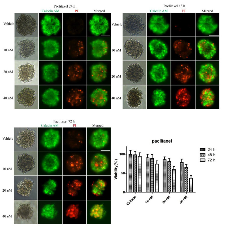
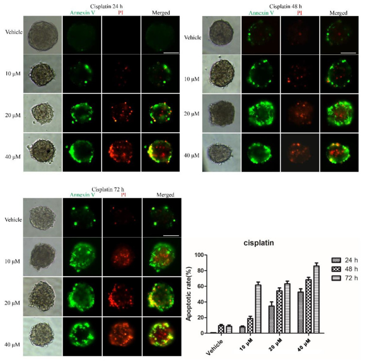
Cell culture is important for the rapid screening of anti-cancer drug candidates, attracting intense interest. Traditional 2D cell culture has been widely utilized in cancer biological research. However, 3D cellular spheroids are able to recapitulate the in vivo microenvironment of tissues or tumors. Thus far, several 3D cell culture methods have been developed, for instance, the hanging drop method, spinner flasks and micropatterned plates. Nevertheless, these methods have been reported to have some disadvantages, for example, medium replacement is inconvenient or causes cellular damage. Here, we report on an easy-to-operate and useful micro-hole culture chip (SimpleDrop) for 3D cellular spheroid formation and culture and drug analysis, which has advantages over the traditional method in terms of its ease of operation, lack of shear force and environmentally friendliness. On this chip, we observed the formation of a 3D spheroid clearly. Three drugs (paclitaxel, cisplatin and methotrexate) were tested by both cell viability assay and drug-induced apoptotic assay. The results show that the three drugs present a similar conclusion: cell viability decreased over time and concentration. Moreover, the apoptotic experiment showed a similar trend to the live/dead cell assay, in that the fraction of the apoptotic and necrotic cells correlated with the concentration and time. All these results prove that our SimpleDrop method is a useful and easy method for the formation of 3D cellular spheroids, which shows its potential for both cell-cell interaction research, tissue engineering and anticancer drug screening.
细胞培养对于抗癌候选药物的快速筛选很重要,因此备受关注。传统的二维细胞培养已广泛应用于癌症生物学研究。然而,三维细胞球体能够重现组织或肿瘤的体内微环境。到目前为止,已经开发了几种三维细胞培养方法,例如悬滴法、旋转瓶和微图案板。然而,据报道这些方法存在一些缺点,例如,培养基更换不方便或会导致细胞损伤。在此,我们报道了一种易于操作且实用的微孔培养芯片(SimpleDrop),用于三维细胞球体的形成、培养和药物分析,该芯片在操作简便性、无剪切力和环境友好性方面优于传统方法。在该芯片上,我们清晰地观察到了三维球体的形成。通过细胞活力测定和药物诱导凋亡测定对三种药物(紫杉醇、顺铂和甲氨蝶呤)进行了测试。结果表明,这三种药物呈现出相似的结论:细胞活力随时间和浓度降低。此外,凋亡实验与活/死细胞测定呈现出相似的趋势,即凋亡和坏死细胞的比例与浓度和时间相关。所有这些结果证明,我们的SimpleDrop方法是一种用于形成三维细胞球体的有用且简便的方法,显示出其在细胞间相互作用研究、组织工程和抗癌药物筛选方面的潜力。