Patil Shankargouda
Department of Maxillofacial Surgery and Diagnostic Sciences, Division of Oral Pathology, College of Dentistry, Jazan University, Jazan 45142, Saudi Arabia.
Curr Issues Mol Biol. 2021 Jun 21;43(1):423-433. doi: 10.3390/cimb43010034.
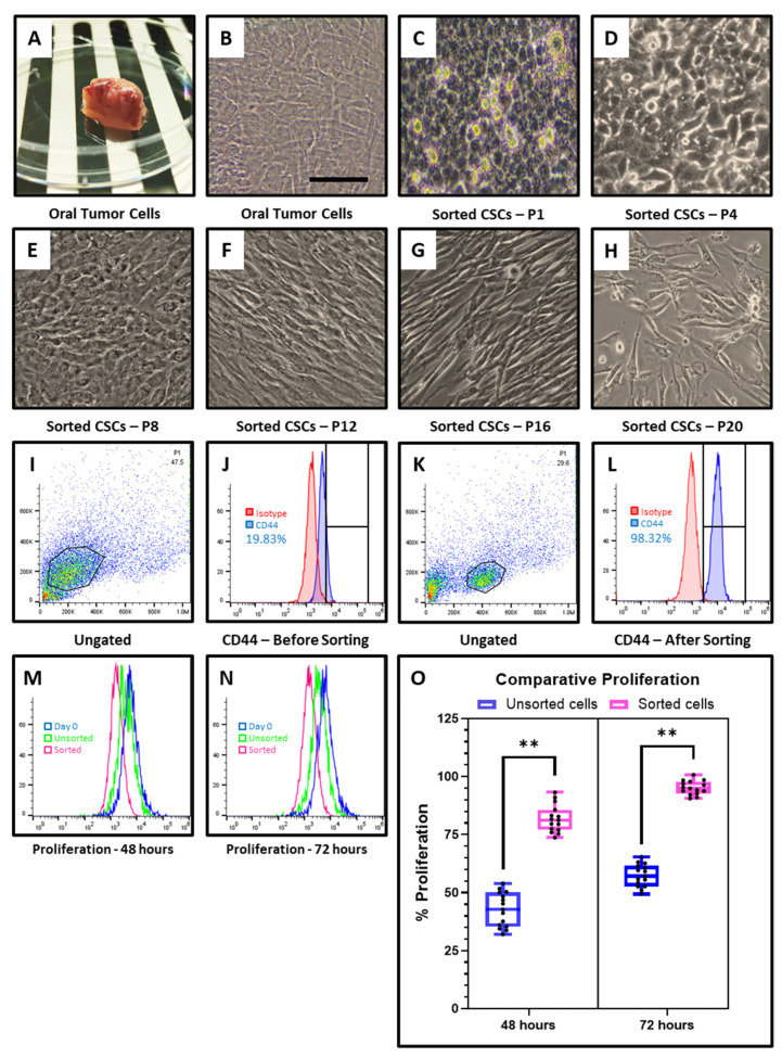
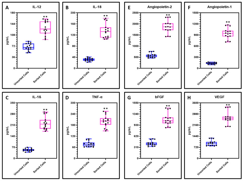
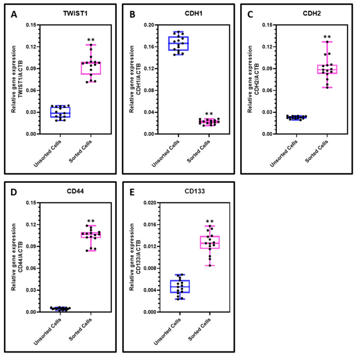
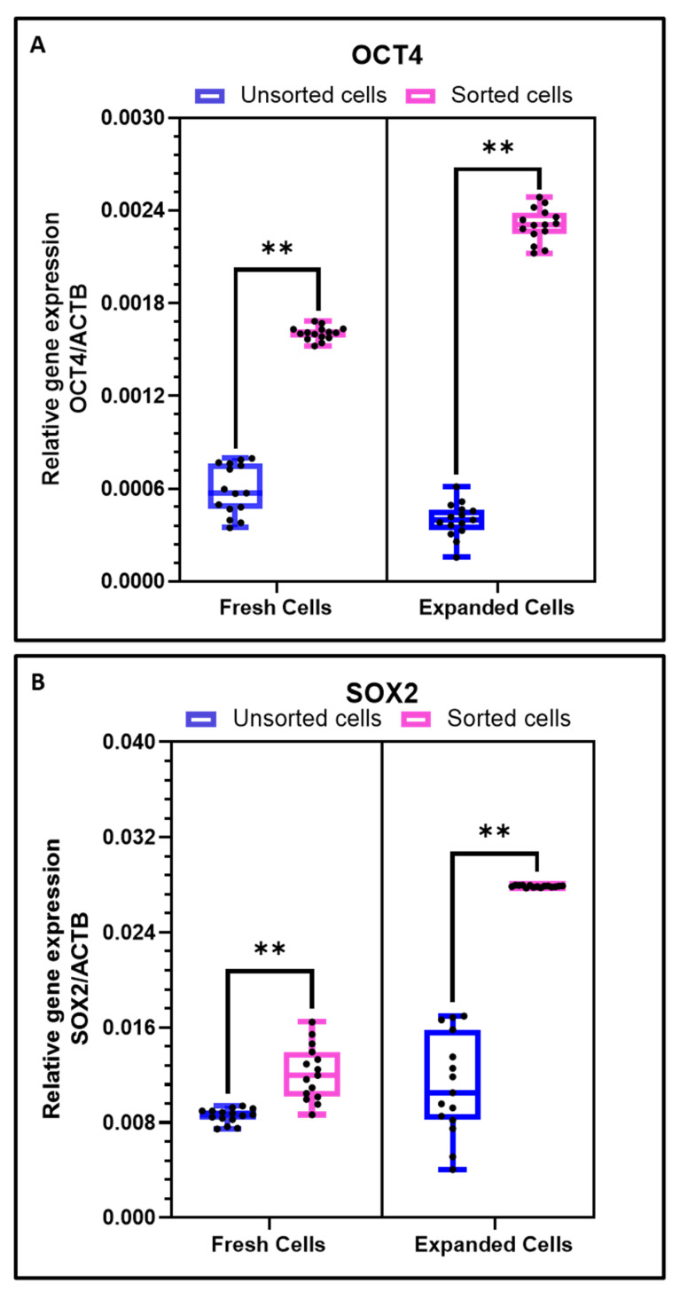
Cancer stem cells (CSCs) have garnered attention with their potential for early diagnosis and prognosis of oral squamous cell carcinoma (OSCC). It is still indistinct whether CSCs are recognized with a specific set of characteristics. The present study aimed to assess the association of CD44 with stemness-related, Epithelial Mesenchymal Transition EMT-related genes and the secretome of the CSCs. The single-cell suspension from primary OSCC tumors was prepared by enzymatic digestion and the cells were cultured in-vitro. The cancer stem cells were isolated by CD44+ selection using magnetic cell-sorting. The expression of CD44, proliferation rate, gene expression of EMT-related transcription factors, stemness markers, cytokine levels and angiogenic factors in both cell population was assessed. The sorted CD44+ cells showed significantly higher proliferation rate than heterogenous population. The CD44 expression was >90% in the sorted cells which was higher than the heterogenous cells. The CD44+ CSCs cells demonstrated significant increased levels of EMT-related genes TWIST1 and CDH2 (-cadherin), CSC-related genes CD44 and CD133 (PROM1), stemness-related genes OCT4, SOX2, inflammatory cytokines IL-1ß, IL-12, IL-18 and TNF-α and angiogenic factors Angiopoietin-1, Angiopoietin-2, bFGF and VEGF while levels of epithelial gene CDH1 (E-cadherin) decreased in comparison to mixed cell population. The genetic and secretome profiling of the CD44+ CSCs could serve as diagnostic and prognostic tools in the treatment of oral cancers.
癌症干细胞(CSCs)因其在口腔鳞状细胞癌(OSCC)早期诊断和预后方面的潜力而备受关注。目前仍不清楚CSCs是否具有一套特定的特征。本研究旨在评估CD44与干性相关、上皮-间质转化(EMT)相关基因以及CSCs分泌组之间的关联。通过酶消化制备原发性OSCC肿瘤的单细胞悬液,并在体外进行细胞培养。使用磁性细胞分选通过CD44 +选择分离癌症干细胞。评估了两种细胞群体中CD44的表达、增殖率、EMT相关转录因子的基因表达、干性标志物、细胞因子水平和血管生成因子。分选后的CD44 +细胞显示出比异质群体显著更高的增殖率。分选细胞中的CD44表达> 90%,高于异质细胞。CD44 + CSCs细胞显示EMT相关基因TWIST1和CDH2(-钙黏蛋白)、CSC相关基因CD44和CD133(PROM1)、干性相关基因OCT4、SOX2、炎性细胞因子IL-1ß、IL-12、IL-18和TNF-α以及血管生成因子血管生成素-1、血管生成素-2、碱性成纤维细胞生长因子(bFGF)和血管内皮生长因子(VEGF)的水平显著升高,而与混合细胞群体相比,上皮基因CDH1(E-钙黏蛋白)水平降低。CD44 + CSCs的基因和分泌组分析可作为口腔癌治疗中的诊断和预后工具。