Zhu Sang, Rong Yan, Kiang Tony K L
Katz Group Centre for Pharmacy and Health Research, Faculty of Pharmacy and Pharmaceutical Sciences, University of Alberta, Edmonton, AB T6G 2E1, Canada.
Pharmaceutics. 2021 Jun 9;13(6):857. doi: 10.3390/pharmaceutics13060857.
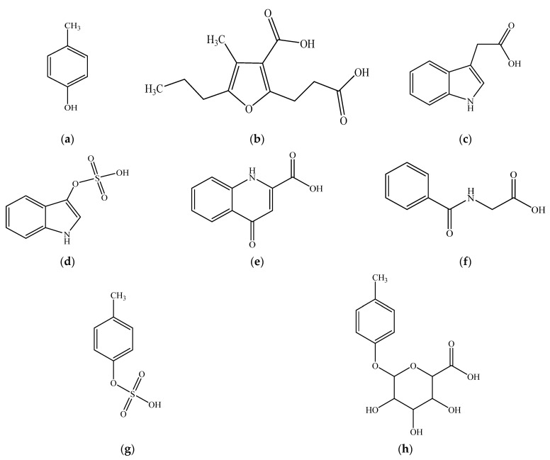
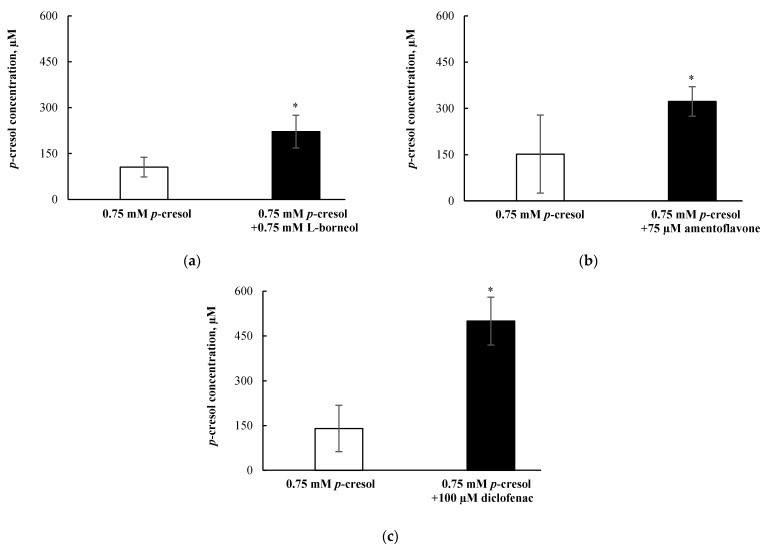
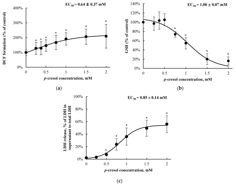
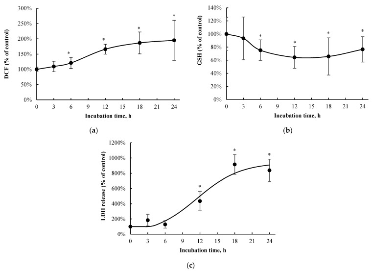
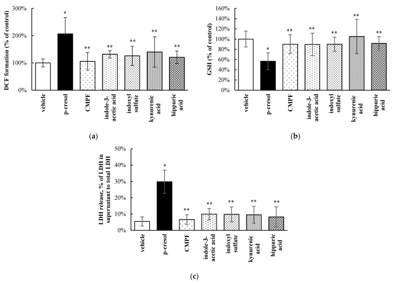
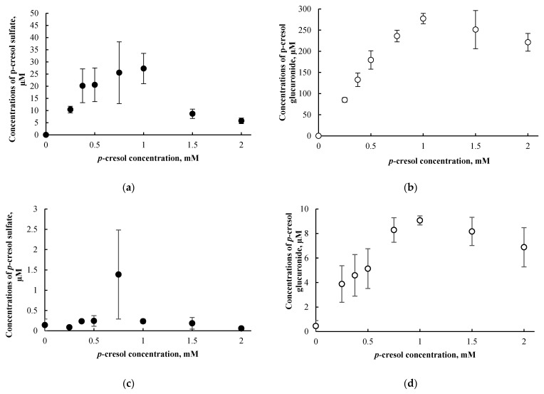
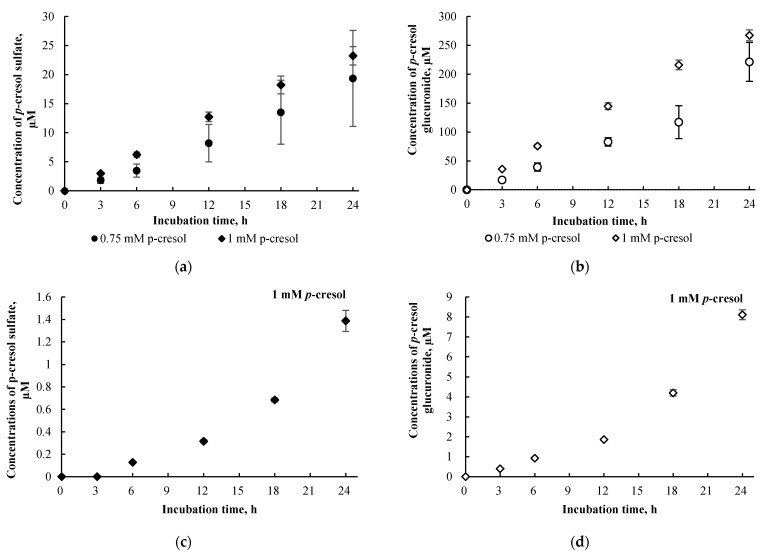
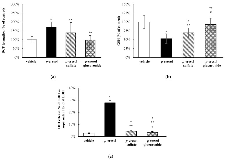
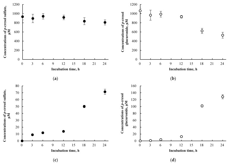
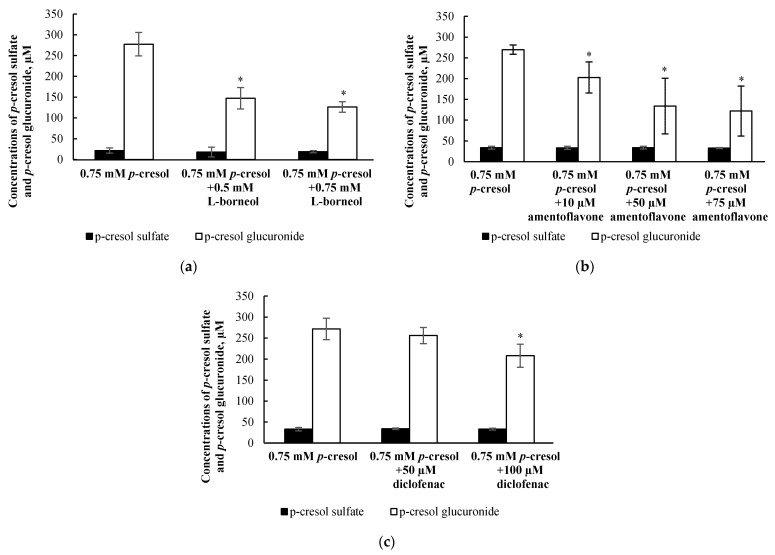
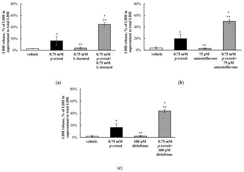
The toxicological effects of -cresol have primarily been attributed to its metabolism products; however, very little human data are available in the key organ (i.e., liver) responsible for the generation of these metabolites. Experiments were conducted in HepaRG cells utilizing the following markers of cellular toxicity: 2'-7'-dichlorofluorescein (DCF; oxidative stress) formation, total cellular glutathione (GSH) concentration, and lactate dehydrogenase (LDH; cellular necrosis) release. Concentrations of -cresol, -cresol sulfate, and -cresol glucuronide were determined using validated assays. -Cresol exposure resulted in concentration- and time-dependent changes in DCF (EC = 0.64 ± 0.37 mM at 24 h of exposure) formation, GSH (EC = 1.00 ± 0.07 mM) concentration, and LDH (EC = 0.85 ± 0.14 mM) release at toxicologically relevant conditions. -Cresol was also relatively more toxic than 3-carboxy-4-methyl-5-propyl-2-furanpropanoic acid, indole-3-acetic acid, indoxyl sulfate, kynurenic acid, and hippuric acid on all markers. Although the exogenous administration of -cresol sulfate and -cresol glucuronide generated high intracellular concentrations of these metabolites, both metabolites were less toxic compared to -cresol at equal-molar conditions. Moreover, -cresol glucuronide was the predominant metabolite generated in situ from -cresol exposure. Selective attenuation of glucuronidation (without affecting -cresol sulfate formation, while increasing -cresol accumulation) using independent chemical inhibitors (i.e., 0.75 mM l-borneol, 75 µM amentoflavone, or 100 µM diclofenac) consistently resulted in further increases in LDH release associated with -cresol exposure (by 28.3 ± 5.3%, 30.0 ± 8.2% or 27.3 ± 6.8%, respectively, compared to -cresol treatment). These novel data indicated that -cresol was a relatively potent toxicant, and that glucuronidation was unlikely to be associated with the manifestation of its toxic effects in HepaRG cells.
对甲酚的毒理学效应主要归因于其代谢产物;然而,在负责生成这些代谢产物的关键器官(即肝脏)中,人类数据非常有限。利用以下细胞毒性标志物在HepaRG细胞中进行了实验:2'-7'-二氯荧光素(DCF;氧化应激)形成、细胞内总谷胱甘肽(GSH)浓度以及乳酸脱氢酶(LDH;细胞坏死)释放。使用经过验证的分析方法测定了对甲酚、硫酸对甲酚和对甲酚葡萄糖醛酸苷的浓度。在毒理学相关条件下,对甲酚暴露导致DCF(暴露24小时时的EC = 0.64 ± 0.37 mM)形成、GSH(EC = 1.00 ± 0.07 mM)浓度和LDH(EC = 0.85 ± 0.14 mM)释放呈现浓度和时间依赖性变化。在所有标志物上,对甲酚的毒性也相对高于3-羧基-4-甲基-5-丙基-2-呋喃丙酸、吲哚-3-乙酸、硫酸吲哚酚、犬尿酸和马尿酸。尽管外源性给予硫酸对甲酚和对甲酚葡萄糖醛酸苷会在细胞内产生高浓度的这些代谢产物,但在等摩尔条件下,这两种代谢产物的毒性均低于对甲酚。此外,对甲酚葡萄糖醛酸苷是对甲酚暴露后原位生成的主要代谢产物。使用独立的化学抑制剂(即0.75 mM l-龙脑、75 µM穗花杉双黄酮或100 µM双氯芬酸)选择性减弱葡萄糖醛酸化(不影响硫酸对甲酚形成,同时增加对甲酚积累)始终导致与对甲酚暴露相关的LDH释放进一步增加(与对甲酚处理相比,分别增加28.3 ± 5.3%、30.0 ± 8.2%或27.3 ± 6.8%)。这些新数据表明,对甲酚是一种相对强效的毒物,并且葡萄糖醛酸化不太可能与其在HepaRG细胞中的毒性作用表现相关。