Loebel Claudia, Kwon Mi Y, Wang Chao, Han Lin, Mauck Robert L, Burdick Jason A
Department of Bioengineering, University of Pennsylvania, 240 Skirkanich Hall, 210 S. 33rd Street, Philadelphia, PA 19104, USA.
School of Biomedical Engineering, Science and Health Systems Drexel University 3141 Chestnut Street, Bossone 718, Philadelphia, PA 19104, USA.
Adv Funct Mater. 2020 Oct 28;30(44). doi: 10.1002/adfm.201909802. Epub 2020 Apr 3.
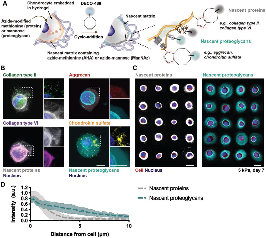
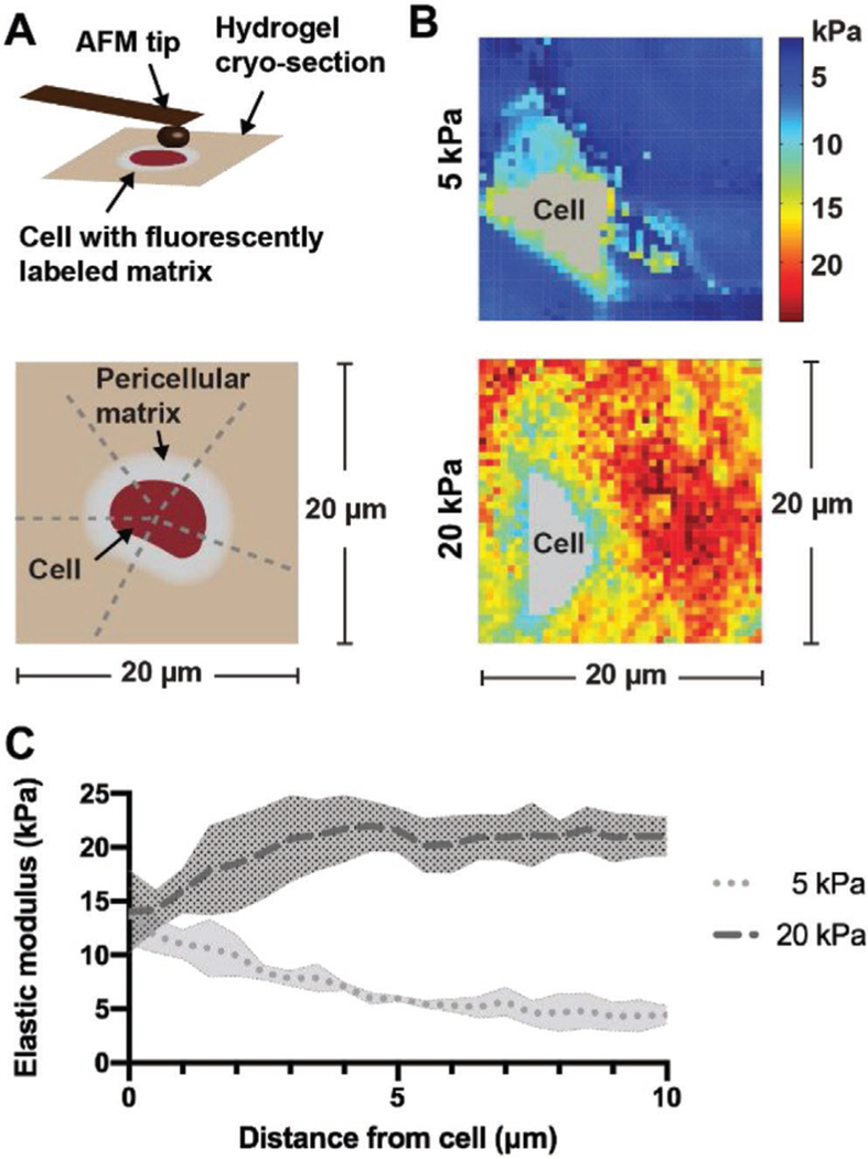
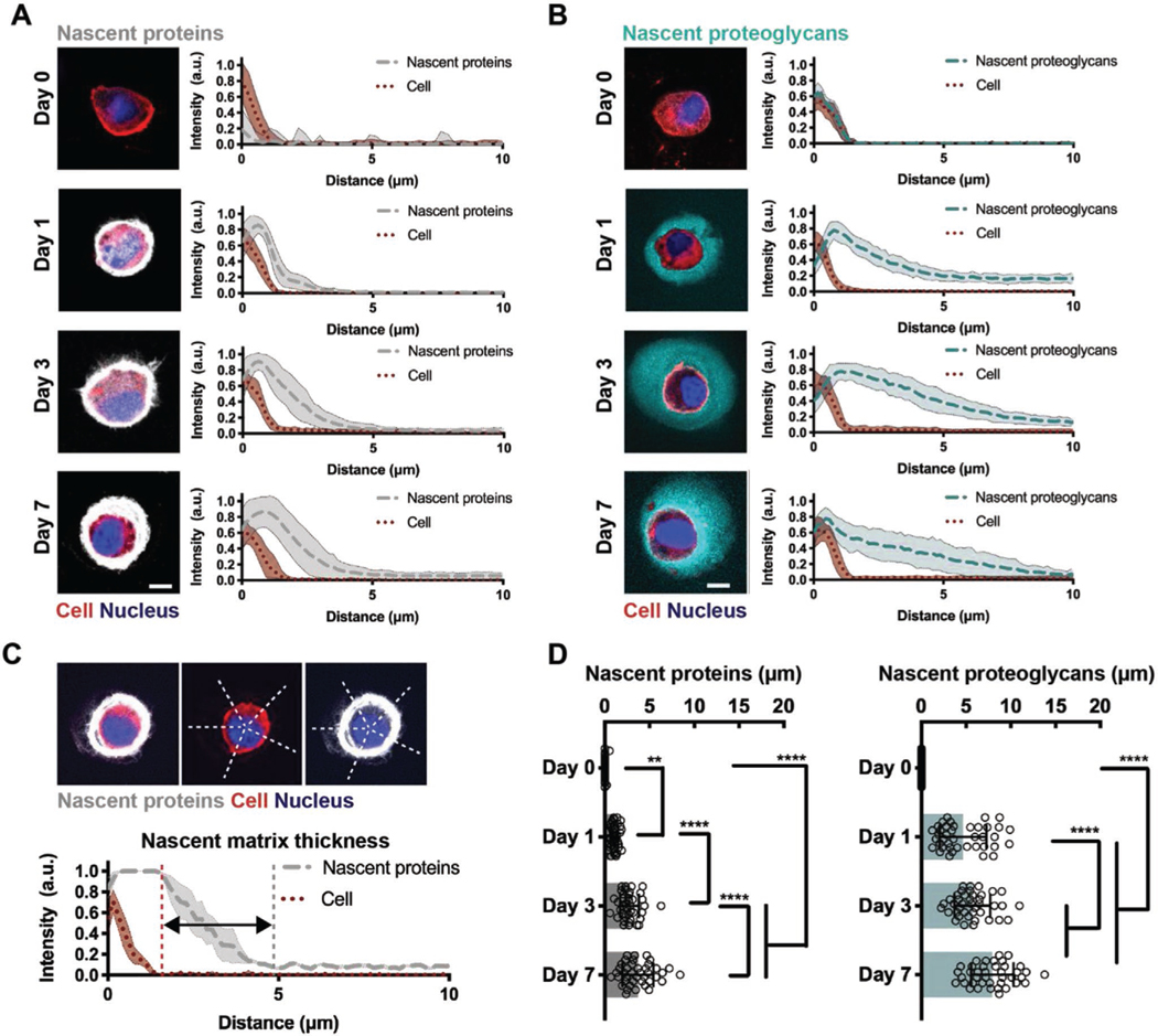
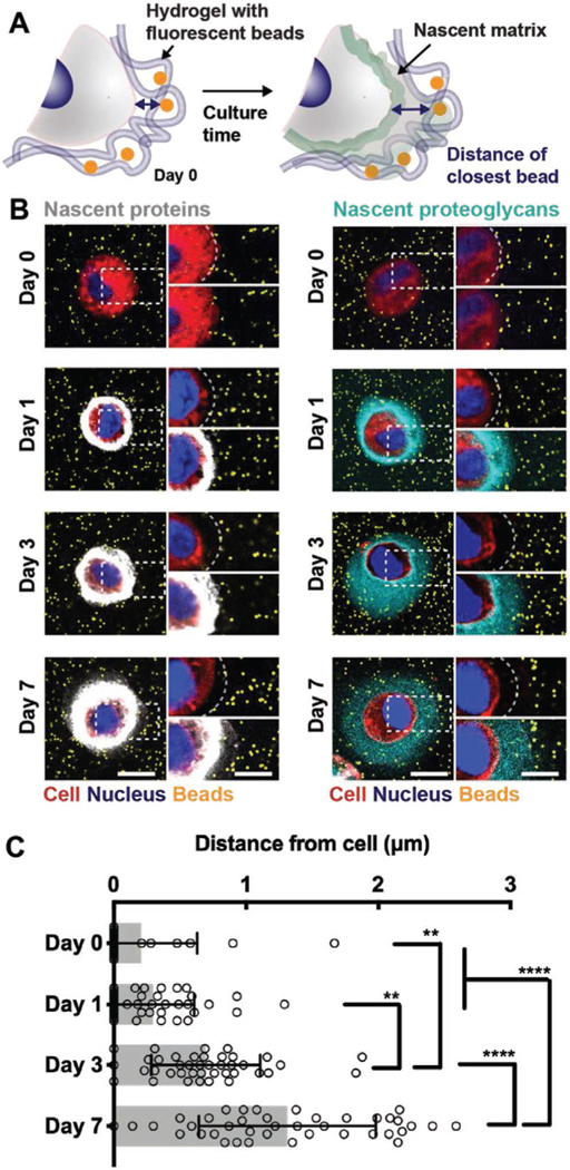
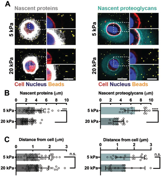
Hydrogels are engineered with biochemical and biophysical signals to recreate aspects of the native microenvironment and to control cellular functions such as differentiation and matrix deposition. This deposited matrix accumulates within the pericellular space and likely affects the interactions between encapsulated cells and the engineered hydrogel; however, there has been little work to study the spatiotemporal evolution of matrix at this interface. To address this, metabolic labeling is employed to visualize the temporal and spatial positioning of nascent proteins and proteoglycans deposited by chondrocytes. Within covalently crosslinked hyaluronic acid hydrogels, chondrocytes deposit nascent proteins and proteoglycans in the pericellular space within 1 d after encapsulation. The accumulation of this matrix, as measured by an increase in matrix thickness during culture, depends on the initial hydrogel crosslink density with decreased thicknesses for more crosslinked hydrogels. Encapsulated fluorescent beads are used to monitor the hydrogel location and indicate that the emerging nascent matrix physically displaces the hydrogel from the cell membrane with extended culture. These findings suggest that secreted matrix increasingly masks the presentation of engineered hydrogel cues and may have implications for the design of hydrogels in tissue engineering and regenerative medicine.
水凝胶通过生化和生物物理信号进行设计,以重现天然微环境的各个方面,并控制细胞功能,如分化和基质沉积。这种沉积的基质在细胞周围空间内积累,并可能影响被包裹细胞与工程水凝胶之间的相互作用;然而,很少有研究致力于探究该界面处基质的时空演变。为了解决这个问题,采用代谢标记来可视化软骨细胞沉积的新生蛋白质和蛋白聚糖的时空定位。在共价交联的透明质酸水凝胶中,软骨细胞在包封后1天内在细胞周围空间沉积新生蛋白质和蛋白聚糖。通过培养过程中基质厚度的增加来衡量,这种基质的积累取决于初始水凝胶的交联密度,交联程度越高的水凝胶厚度越小。使用包封的荧光珠来监测水凝胶的位置,并表明随着培养时间延长,新出现的新生基质会将水凝胶从细胞膜处物理性地推开。这些发现表明,分泌的基质越来越多地掩盖了工程水凝胶信号的呈现,这可能对组织工程和再生医学中水凝胶的设计产生影响。