García-Varela Lara, Rodríguez-Pérez Manuel, Custodia Antía, Moraga-Amaro Rodrigo, Colabufo Nicola A, Aguiar Pablo, Sobrino Tomás, Dierckx Rudi A J O, van Waarde Aren, Elsinga Philip H, Luurtsema Gert
Department of Nuclear Medicine and Molecular Imaging, University of Groningen, University Medical Center Groningen, Hanzeplein 1, P.O. Box 30001, 9713 GZ Groningen, The Netherlands.
Clinical Neurosciences Research Laboratory, Health Research Institute of Santiago de Compostela (IDIS), 15706 Santiago de Compostela, Spain.
Mol Pharm. 2021 Aug 2;18(8):3073-3085. doi: 10.1021/acs.molpharmaceut.1c00302. Epub 2021 Jul 6.
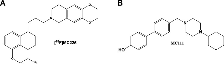
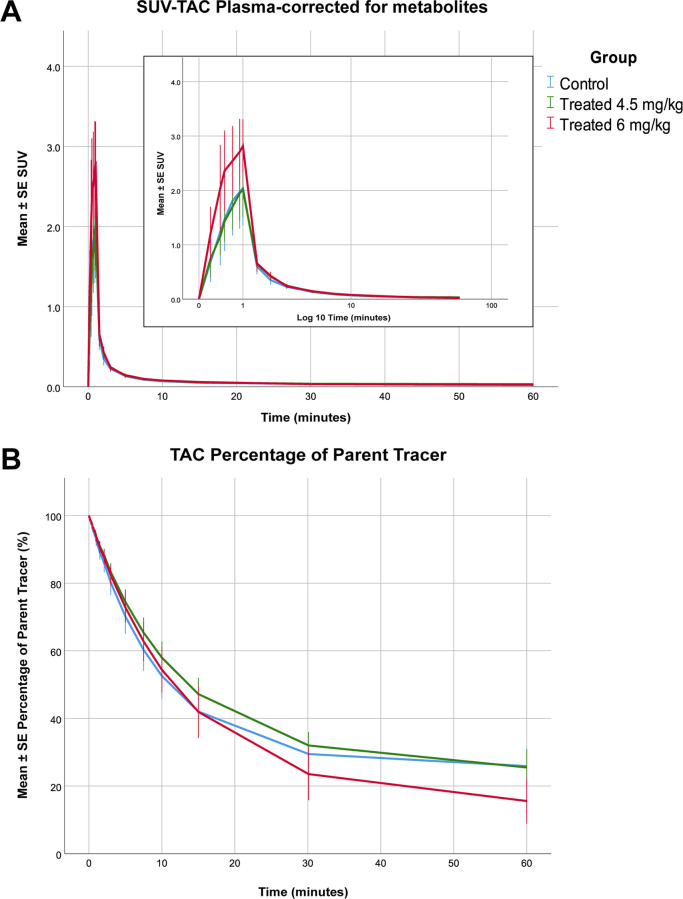
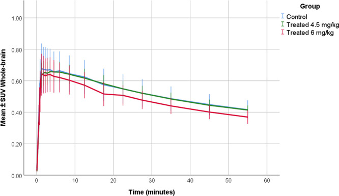
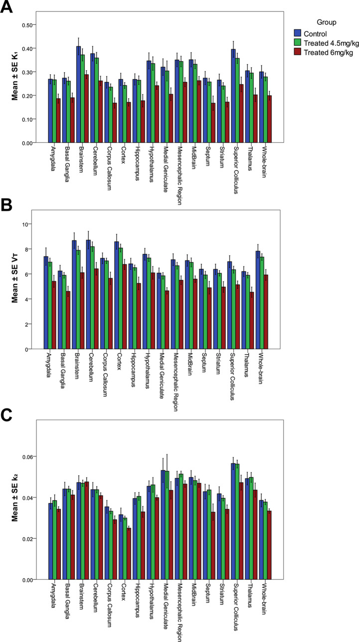
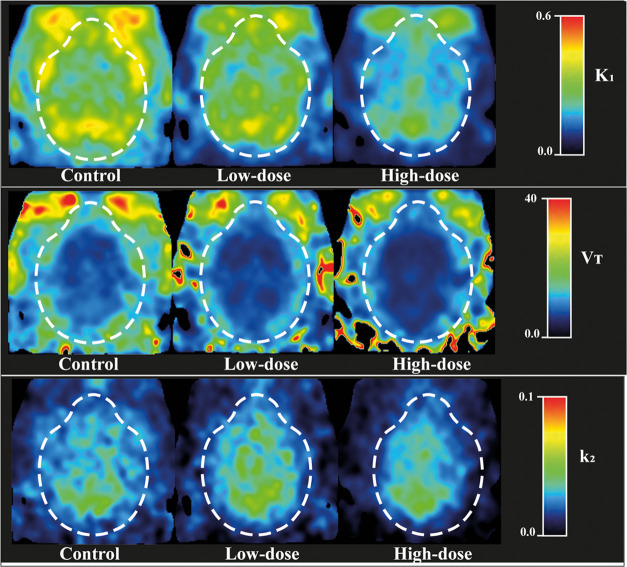
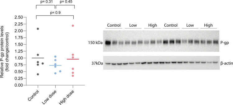
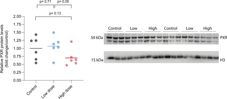
P-Glycoprotein (P-gp) is an efflux pump located at the blood-brain barrier (BBB) that contributes to the protection of the central nervous system by transporting neurotoxic compounds out of the brain. A decline in P-gp function has been related to the pathogenesis of neurodegenerative diseases. P-gp inducers can increase the P-gp function and are considered as potential candidates for the treatment of such disorders. The P-gp inducer MC111 increased P-gp expression and function in SW480 human colon adenocarcinoma and colo-320 cells, respectively. Our study aims to evaluate the P-gp inducing effect of MC111 in the whole brain , using the P-gp tracer [F]MC225 and positron emission tomography (PET). Eighteen Wistar rats were treated with either vehicle solution, 4.5 mg/kg of MC111 (low-dose group), or 6 mg/kg of MC111 (high-dose group). Animals underwent a 60 min dynamic PET scan with arterial-blood sampling, 24 h after treatment with the inducer. Data were analyzed using the 1-tissue-compartment model and metabolite-corrected plasma as the input function. Model parameters such as the influx constant () and volume of distribution () were calculated, which reflect the P-gp function. P-gp and pregnane xenobiotic receptor (PXR) expression levels of the whole brain were assessed using western blot. The administration of MC111 decreased and of [F]MC225 in the whole brain and all of the selected brain regions. In the high-dose group, whole-brain was decreased by 34% (-high-dose = 0.20 ± 0.02 vs -control = 0.30 ± 0.02; < 0.001) and in the low-dose group by 7% (-low-dose = 0.28 ± 0.02 vs -control = 0.30 ± 0.02; = 0.42) compared to controls. Whole-brain was decreased by 25% in the high-dose group (-high-dose = 5.92 ± 0.41 vs -control = 7.82 ± 0.38; < 0.001) and by 6% in the low-dose group (-low-dose = 7.35 ± 0.38 vs -control = 7.82 ± 0.37; = 0.38) compared to controls. values did not vary after treatment. The treatment did not affect the metabolism of [F]MC225. Western blot studies using the whole-brain tissue did not detect changes in the P-gp expression, however, preliminary results using isolated brain capillaries found an increasing trend up to 37% in treated rats. The decrease in and values after treatment with the inducer indicates an increase in the P-gp functionality at the BBB of treated rats. Moreover, preliminary results using brain endothelial cells also sustained the increase in the P-gp expression. In conclusion, the results verify that MC111 induces P-gp expression and function at the BBB in rats. An increasing trend regarding the P-gp expression levels is found using western blot and an increased P-gp function is confirmed with [F]MC225 and PET.
P-糖蛋白(P-gp)是一种位于血脑屏障(BBB)的外排泵,它通过将神经毒性化合物转运出脑来保护中枢神经系统。P-gp功能的下降与神经退行性疾病的发病机制有关。P-gp诱导剂可增强P-gp功能,被认为是治疗此类疾病的潜在候选药物。P-gp诱导剂MC111分别增加了SW480人结肠腺癌和colo-320细胞中的P-gp表达和功能。我们的研究旨在使用P-gp示踪剂[F]MC225和正电子发射断层扫描(PET)评估MC111在全脑中的P-gp诱导作用。18只Wistar大鼠分别接受赋形剂溶液、4.5mg/kg的MC111(低剂量组)或6mg/kg的MC111(高剂量组)处理。在用诱导剂处理24小时后,动物进行了60分钟的动态PET扫描并采集动脉血样。使用1组织室模型并以代谢物校正的血浆作为输入函数对数据进行分析。计算了反映P-gp功能的模型参数,如流入常数()和分布容积()。使用蛋白质印迹法评估全脑的P-gp和孕烷异生素受体(PXR)表达水平。MC111的给药降低了全脑和所有选定脑区中[F]MC225的和。在高剂量组中,全脑降低了34%(-高剂量=0.20±0.02 vs -对照=0.30±0.02;<0.001),在低剂量组中与对照组相比降低了7%(-低剂量=0.28±0.02 vs -对照=0.30±0.02;=0.42)。高剂量组中全脑降低了25%(-高剂量=5.92±0.41 vs -对照=7.82±0.38;<0.001),低剂量组中与对照组相比降低了6%(-低剂量=7.35±0.38 vs -对照=7.82±0.37;=0.38)。处理后值没有变化。该处理不影响[F]MC225的代谢。使用全脑组织的蛋白质印迹研究未检测到P-gp表达的变化,然而,使用分离的脑毛细血管的初步结果发现,处理后的大鼠中P-gp表达有高达37%的增加趋势。诱导剂处理后和值的降低表明处理后大鼠血脑屏障处的P-gp功能增强。此外,使用脑内皮细胞的初步结果也证实了P-gp表达的增加。总之,结果证实MC111在大鼠血脑屏障处诱导P-gp表达和功能。使用蛋白质印迹法发现P-gp表达水平有增加趋势,并用[F]MC225和PET证实了P-gp功能增强。