• 文献检索
  • 文档翻译
  • 深度研究
  • 学术资讯
  • Suppr Zotero 插件Zotero 插件
  • 邀请有礼
  • 套餐&价格
  • 历史记录
应用&插件
Suppr Zotero 插件Zotero 插件浏览器插件Mac 客户端Windows 客户端微信小程序
定价
高级版会员购买积分包购买API积分包
服务
文献检索文档翻译深度研究API 文档MCP 服务
关于我们
关于 Suppr公司介绍联系我们用户协议隐私条款
关注我们

Suppr 超能文献

核心技术专利:CN118964589B侵权必究
粤ICP备2023148730 号-1Suppr @ 2026

文献检索

告别复杂PubMed语法,用中文像聊天一样搜索,搜遍4000万医学文献。AI智能推荐,让科研检索更轻松。

立即免费搜索

文件翻译

保留排版,准确专业,支持PDF/Word/PPT等文件格式,支持 12+语言互译。

免费翻译文档

深度研究

AI帮你快速写综述,25分钟生成高质量综述,智能提取关键信息,辅助科研写作。

立即免费体验

磁共振成像放射组学预测术前腋窝淋巴结转移以支持手术决策,并与浸润性乳腺癌的肿瘤微环境相关:一项机器学习、多中心研究。

Magnetic resonance imaging radiomics predicts preoperative axillary lymph node metastasis to support surgical decisions and is associated with tumor microenvironment in invasive breast cancer: A machine learning, multicenter study.

机构信息

Guangdong Provincial Key Laboratory of Malignant Tumor Epigenetics and Gene Regulation, Department of Medical Oncology, Breast Tumor Centre, Phase I Clinical Trial Centre, Sun Yat-sen Memorial Hospital, Sun Yat-sen University, Guangzhou, China.

Department of Breast Surgery, Tungwah Hospital, Sun Yat-sen University, Dongguan, China.

出版信息

EBioMedicine. 2021 Jul;69:103460. doi: 10.1016/j.ebiom.2021.103460. Epub 2021 Jul 4.

DOI:10.1016/j.ebiom.2021.103460
PMID:34233259
原文链接:https://pmc.ncbi.nlm.nih.gov/articles/PMC8261009/
Abstract

BACKGROUND

in current clinical practice, the standard evaluation for axillary lymph node (ALN) status in breast cancer has a low efficiency and is based on an invasive procedure that causes operative-associated complications in many patients. Therefore, we aimed to use machine learning techniques to develop an efficient preoperative magnetic resonance imaging (MRI) radiomics evaluation approach of ALN status and explore the association between radiomics and the tumor microenvironment in patients with early-stage invasive breast cancer.

METHODS

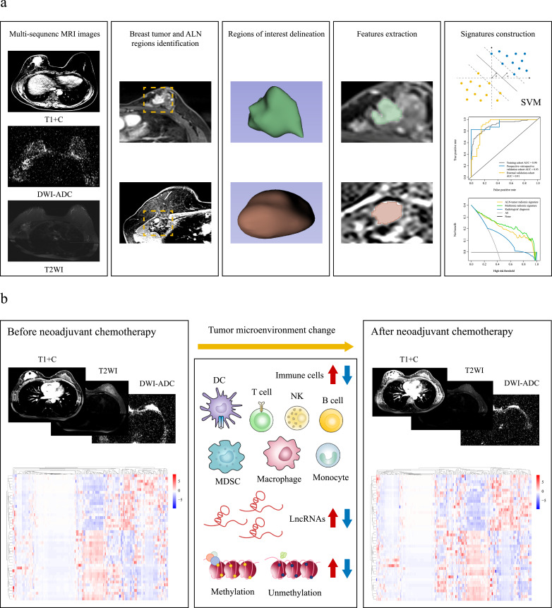
in this retrospective multicenter study, three independent cohorts of patients with breast cancer (n = 1,088) were used to develop and validate signatures predictive of ALN status. After applying the machine learning random forest algorithm to select the key preoperative MRI radiomic features, we used ALN and tumor radiomic features to develop the ALN-tumor radiomic signature for ALN status prediction by the support vector machine algorithm in 803 patients with breast cancer from Sun Yat-sen Memorial Hospital and Sun Yat-sen University Cancer Center (training cohort). By combining ALN and tumor radiomic features with corresponding clinicopathologic information, the multiomic signature was constructed in the training cohort. Next, the external validation cohort (n = 179) of patients from Shunde Hospital of Southern Medical University and Tungwah Hospital of Sun Yat-Sen University, and the prospective-retrospective validation cohort (n = 106) of patients treated with neoadjuvant chemotherapy in prospective phase 3 trials [NCT01503905], were included to evaluate the predictive value of the two signatures, and their predictive performance was assessed by the area under operating characteristic curve (AUC). This study was registered with ClinicalTrials.gov, number NCT04003558.

FINDINGS

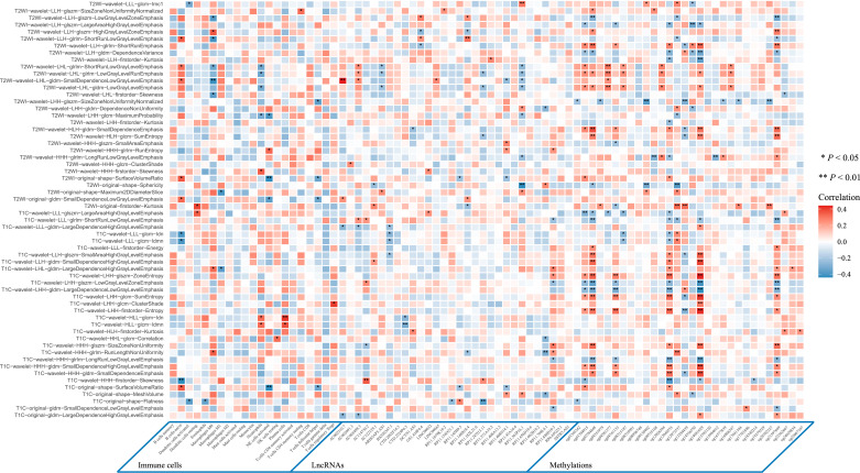
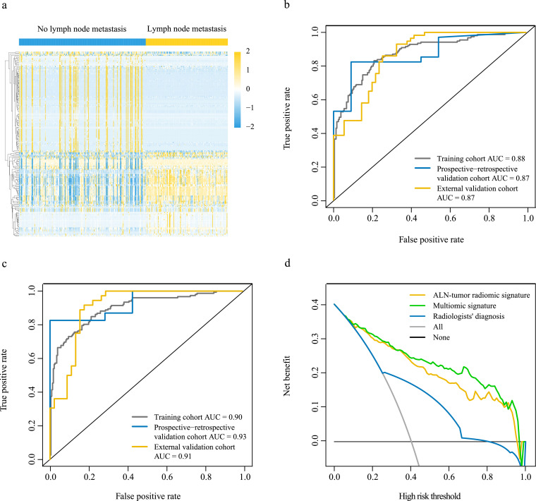
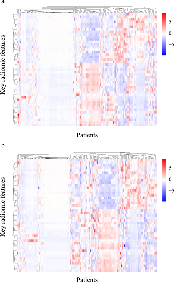
the ALN-tumor radiomic signature for ALN status prediction comprising ALN and tumor radiomic features showed a high prediction quality with AUC of 0·88 in the training cohort, 0·87 in the external validation cohort, and 0·87 in the prospective-retrospective validation cohort. The multiomic signature incorporating tumor and lymph node MRI radiomics, clinical and pathologic characteristics, and molecular subtypes achieved better performance for ALN status prediction with AUCs of 0·90, 0·91, and 0·93 in the training cohort, the external validation cohort, and the prospective-retrospective validation cohort, respectively. Among patients who underwent neoadjuvant chemotherapy in the prospective-retrospective validation cohort, there were significant differences in the key radiomic features before and after neoadjuvant chemotherapy, especially in the gray-level dependence matrix features. Furthermore, there was an association between MRI radiomics and tumor microenvironment features including immune cells, long non-coding RNAs, and types of methylated sites. Interpretation this study presented a multiomic signature that could be preoperatively and conveniently used for identifying patients with ALN metastasis in early-stage invasive breast cancer. The multiomic signature exhibited powerful predictive ability and showed the prospect of extended application to tailor surgical management. Besides, significant changes in key radiomic features after neoadjuvant chemotherapy may be explained by changes in the tumor microenvironment, and the association between MRI radiomic features and tumor microenvironment features may reveal the potential biological underpinning of MRI radiomics.

FUNDING

No funding.

摘要

背景

在当前的临床实践中,腋窝淋巴结(ALN)状态的标准评估效率低下,且基于一种会在许多患者中引起手术相关并发症的侵入性操作。因此,我们旨在使用机器学习技术开发一种有效的术前磁共振成像(MRI)放射组学 ALN 状态评估方法,并探讨放射组学与早期浸润性乳腺癌患者肿瘤微环境之间的关联。

方法

本回顾性多中心研究纳入了三批乳腺癌患者队列(n=1088),用于开发和验证预测 ALN 状态的特征。我们应用机器学习随机森林算法选择关键的术前 MRI 放射组学特征,然后使用 ALN 和肿瘤放射组学特征,通过支持向量机算法,在中山大学孙逸仙纪念医院和中山大学肿瘤防治中心的 803 例乳腺癌患者(训练队列)中开发用于预测 ALN 状态的 ALN-肿瘤放射组学特征。通过结合 ALN 和肿瘤放射组学特征与相应的临床病理信息,我们在训练队列中构建了多组学特征。然后,纳入南方医科大学顺德医院和中山大学孙逸仙纪念医院的患者外部验证队列(n=179),以及前瞻性、回顾性验证队列(n=106),该队列患者在前瞻性 III 期临床试验[ NCT01503905]中接受新辅助化疗,以评估这两个特征的预测价值,并通过接受者操作特征曲线下面积(AUC)评估其预测性能。本研究在 ClinicalTrials.gov 上注册,编号为 NCT04003558。

结果

ALN-肿瘤放射组学特征预测 ALN 状态的特征,包括 ALN 和肿瘤放射组学特征,在训练队列中具有 0.88 的高预测质量,在外部验证队列中为 0.87,在前瞻性、回顾性验证队列中为 0.87。纳入肿瘤和淋巴结 MRI 放射组学、临床病理特征和分子亚型的多组学特征,在训练队列、外部验证队列和前瞻性、回顾性验证队列中的 ALN 状态预测中,获得了更好的性能,AUC 分别为 0.90、0.91 和 0.93。在前瞻性、回顾性验证队列中接受新辅助化疗的患者中,新辅助化疗前后关键放射组学特征存在显著差异,尤其是灰度依赖矩阵特征。此外,MRI 放射组学与包括免疫细胞、长链非编码 RNA 和甲基化位点类型在内的肿瘤微环境特征之间存在关联。

结论

本研究提出了一种多组学特征,可以在术前方便地用于识别早期浸润性乳腺癌中的 ALN 转移患者。多组学特征表现出强大的预测能力,并显示出在制定手术管理方案方面的扩展应用前景。此外,新辅助化疗后关键放射组学特征的变化可能是肿瘤微环境变化的结果,MRI 放射组学特征与肿瘤微环境特征之间的关联可能揭示了 MRI 放射组学的潜在生物学基础。

资金

无。

https://cdn.ncbi.nlm.nih.gov/pmc/blobs/450c/8261009/542dc071c31c/gr5.jpg
https://cdn.ncbi.nlm.nih.gov/pmc/blobs/450c/8261009/85c05323f7ef/gr1.jpg
https://cdn.ncbi.nlm.nih.gov/pmc/blobs/450c/8261009/4420105ad4e3/gr2.jpg
https://cdn.ncbi.nlm.nih.gov/pmc/blobs/450c/8261009/a535a8c9bbe0/gr3.jpg
https://cdn.ncbi.nlm.nih.gov/pmc/blobs/450c/8261009/eb9fce409126/gr4.jpg
https://cdn.ncbi.nlm.nih.gov/pmc/blobs/450c/8261009/542dc071c31c/gr5.jpg

相似文献

1
Magnetic resonance imaging radiomics predicts preoperative axillary lymph node metastasis to support surgical decisions and is associated with tumor microenvironment in invasive breast cancer: A machine learning, multicenter study.磁共振成像放射组学预测术前腋窝淋巴结转移以支持手术决策,并与浸润性乳腺癌的肿瘤微环境相关:一项机器学习、多中心研究。
EBioMedicine. 2021 Jul;69:103460. doi: 10.1016/j.ebiom.2021.103460. Epub 2021 Jul 4.
2
Development and Validation of a Preoperative Magnetic Resonance Imaging Radiomics-Based Signature to Predict Axillary Lymph Node Metastasis and Disease-Free Survival in Patients With Early-Stage Breast Cancer.基于术前磁共振成像放射组学的signature 模型:预测早期乳腺癌患者腋窝淋巴结转移和无病生存的研究
JAMA Netw Open. 2020 Dec 1;3(12):e2028086. doi: 10.1001/jamanetworkopen.2020.28086.
3
Preoperative prediction of lymphovascular invasion in invasive breast cancer with dynamic contrast-enhanced-MRI-based radiomics.基于动态对比增强 MRI 的放射组学术前预测浸润性乳腺癌的淋巴管血管侵犯。
J Magn Reson Imaging. 2019 Sep;50(3):847-857. doi: 10.1002/jmri.26688. Epub 2019 Feb 17.
4
Multi-modality radiomics model predicts axillary lymph node metastasis of breast cancer using MRI and mammography.多模态放射组学模型利用 MRI 和乳腺 X 线摄影预测乳腺癌腋窝淋巴结转移。
Eur Radiol. 2024 Sep;34(9):6121-6131. doi: 10.1007/s00330-024-10638-2. Epub 2024 Feb 10.
5
Preoperative prediction of axillary sentinel lymph node burden with multiparametric MRI-based radiomics nomogram in early-stage breast cancer.基于多参数 MRI 放射组学列线图预测早期乳腺癌腋窝前哨淋巴结负荷
Eur Radiol. 2021 Aug;31(8):5924-5939. doi: 10.1007/s00330-020-07674-z. Epub 2021 Feb 10.
6
Non-invasive prediction model of axillary lymph node status in patients with early-stage breast cancer: a feasibility study based on dynamic contrast-enhanced-MRI radiomics.基于动态对比增强 MRI 放射组学的早期乳腺癌患者腋窝淋巴结状态的无创预测模型:一项可行性研究。
Br J Radiol. 2024 Feb 2;97(1154):439-450. doi: 10.1093/bjr/tqad034.
7
Machine Learning Model for Predicting Axillary Lymph Node Metastasis in Clinically Node Positive Breast Cancer Based on Peritumoral Ultrasound Radiomics and SHAP Feature Analysis.基于瘤周超声影像组学和SHAP特征分析的临床淋巴结阳性乳腺癌腋窝淋巴结转移预测机器学习模型
J Ultrasound Med. 2024 Sep;43(9):1611-1625. doi: 10.1002/jum.16483. Epub 2024 May 29.
8
Non-invasive prediction of axillary lymph node dissection exemption in breast cancer patients post-neoadjuvant therapy: A radiomics and deep learning analysis on longitudinal DCE-MRI data.新辅助治疗后乳腺癌患者腋窝淋巴结清扫豁免的无创预测:纵向 DCE-MRI 数据的放射组学和深度学习分析。
Breast. 2024 Oct;77:103786. doi: 10.1016/j.breast.2024.103786. Epub 2024 Aug 9.
9
A delta-radiomic lymph node model using dynamic contrast enhanced MRI for the early prediction of axillary response after neoadjuvant chemotherapy in breast cancer patients.基于动态对比增强 MRI 的 delta 放射组学淋巴结模型预测乳腺癌新辅助化疗后腋窝早期疗效。
BMC Cancer. 2023 Jan 5;23(1):15. doi: 10.1186/s12885-022-10496-5.
10
A two-center study of a combined nomogram based on mammography and MRI to predict ALN metastasis in breast cancer.基于乳腺 X 线摄影和 MRI 的联合列线图预测乳腺癌前哨淋巴结转移的两中心研究。
Magn Reson Imaging. 2024 Jul;110:128-137. doi: 10.1016/j.mri.2024.04.019. Epub 2024 Apr 15.

引用本文的文献

1
Development of a Machine Learning Model Integrating Pathomics and Clinical Data to Predict Axillary Lymph Node Metastasis in Breast Cancer: A Two-Center Study.整合病理组学和临床数据的机器学习模型用于预测乳腺癌腋窝淋巴结转移的研究:一项双中心研究
Cancer Rep (Hoboken). 2025 Sep;8(9):e70302. doi: 10.1002/cnr2.70302.
2
Exploring the impact of high rosuvastatin plasma exposure on new-onset diabetes mellitus: insights from machine learning-based prediction.探索瑞舒伐他汀高血浆暴露量对新发糖尿病的影响:基于机器学习预测的见解
Eur J Med Res. 2025 Aug 25;30(1):796. doi: 10.1186/s40001-025-03029-w.
3
Development and Validation of a Computed Tomography-based Radiomics Nomogram for Diagnosing Cervical Lymph Node Metastasis in Oropharyngeal Squamous Cell Carcinomas.
基于计算机断层扫描的影像组学列线图在诊断口咽鳞状细胞癌颈部淋巴结转移中的开发与验证
Adv Radiat Oncol. 2025 Jul 1;10(9):101844. doi: 10.1016/j.adro.2025.101844. eCollection 2025 Sep.
4
Development and validation of an F-FDG PET/CT radiomic nomogram for predicting axillary lymph-node status after neoadjuvant chemotherapy for breast cancer: a multicenter study.用于预测乳腺癌新辅助化疗后腋窝淋巴结状态的F-FDG PET/CT影像组学列线图的开发与验证:一项多中心研究
Ann Nucl Med. 2025 Aug 17. doi: 10.1007/s12149-025-02099-4.
5
Using baseline MRI radiomics to predict the tumor shrinkage patterns in HR-Positive, HER2-Negative Breast Cancer.利用基线MRI影像组学预测激素受体阳性、人表皮生长因子受体2阴性乳腺癌的肿瘤缩小模式。
Front Oncol. 2025 Jul 30;15:1539644. doi: 10.3389/fonc.2025.1539644. eCollection 2025.
6
An explainable predictive machine learning model for axillary lymph node metastasis in breast cancer based on multimodal data: A retrospective single-center study.基于多模态数据的可解释性乳腺癌腋窝淋巴结转移预测机器学习模型:一项回顾性单中心研究。
J Transl Med. 2025 Aug 11;23(1):892. doi: 10.1186/s12967-025-06686-x.
7
Automatic recognition of adrenal incidentalomas using a two-stage cascade network: a multicenter study.使用两阶段级联网络自动识别肾上腺偶发瘤:一项多中心研究
Ann Med. 2025 Dec;57(1):2540596. doi: 10.1080/07853890.2025.2540596. Epub 2025 Aug 7.
8
Development and validation of MRI-based radiomics model for clinical symptom stratification of extrinsic adenomyosis.基于MRI的外源性子宫腺肌病临床症状分层的影像组学模型的开发与验证
Ann Med. 2025 Dec;57(1):2534521. doi: 10.1080/07853890.2025.2534521. Epub 2025 Jul 25.
9
Integrative radiomics of intra- and peri-tumoral features for enhanced risk prediction in thymic tumors: a multimodal analysis of tumor microenvironment contributions.整合瘤内和瘤周特征的放射组学以增强胸腺瘤风险预测:肿瘤微环境贡献的多模态分析
BMC Med Imaging. 2025 Jul 17;25(1):286. doi: 10.1186/s12880-025-01790-2.
10
Deep learning on routine full-breast mammograms enhances lymph node metastasis prediction in early breast cancer.基于常规全乳钼靶X线摄影的深度学习可提高早期乳腺癌淋巴结转移的预测能力。
NPJ Digit Med. 2025 Jul 10;8(1):425. doi: 10.1038/s41746-025-01831-8.