• 文献检索
  • 文档翻译
  • 深度研究
  • 学术资讯
  • Suppr Zotero 插件Zotero 插件
  • 邀请有礼
  • 套餐&价格
  • 历史记录
应用&插件
Suppr Zotero 插件Zotero 插件浏览器插件Mac 客户端Windows 客户端微信小程序
定价
高级版会员购买积分包购买API积分包
服务
文献检索文档翻译深度研究API 文档MCP 服务
关于我们
关于 Suppr公司介绍联系我们用户协议隐私条款
关注我们

Suppr 超能文献

核心技术专利:CN118964589B侵权必究
粤ICP备2023148730 号-1Suppr @ 2026

文献检索

告别复杂PubMed语法,用中文像聊天一样搜索,搜遍4000万医学文献。AI智能推荐,让科研检索更轻松。

立即免费搜索

文件翻译

保留排版,准确专业,支持PDF/Word/PPT等文件格式,支持 12+语言互译。

免费翻译文档

深度研究

AI帮你快速写综述,25分钟生成高质量综述,智能提取关键信息,辅助科研写作。

立即免费体验

帕金森病患者迷走神经横截面积——一项超声病例对照研究

Vagus Nerve Cross-Sectional Area in Patients With Parkinson's Disease-An Ultrasound Case-Control Study.

作者信息

Horsager Jacob, Walter Uwe, Fedorova Tatyana D, Andersen Katrine B, Skjærbæk Casper, Knudsen Karoline, Okkels Niels, von Weitzel-Mudersbach Paul, Dyrskog Stig Eric, Bergholt Bo, Borghammer Per

机构信息

Department of Nuclear Medicine and PET, Aarhus University Hospital, Aarhus, Denmark.

Department of Neurology, Rostock University, Rostock, Germany.

出版信息

Front Neurol. 2021 Jun 22;12:681413. doi: 10.3389/fneur.2021.681413. eCollection 2021.

DOI:10.3389/fneur.2021.681413
PMID:34239497
原文链接:https://pmc.ncbi.nlm.nih.gov/articles/PMC8258145/
Abstract

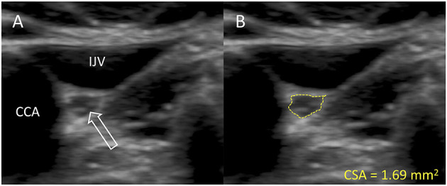
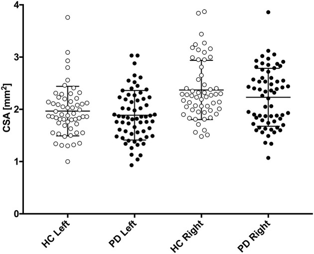
Vagal parasympathetic neurons are prone to degeneration in Parkinson's disease (PD). High-resolution ultrasound can precisely estimate the cross-sectional (CSA) area of peripheral nerves. Here, we tested the hypothesis that vagus CSA is reduced in PD. We included 56 healthy controls (HCs) and 63 patients with PD. Using a high-end ultrasound system equipped with a high-frequency transducer, five images were obtained of each nerve. The hypoechoic neuronal tissue was delineated offline with dedicated software and the CSA extracted. In the initial PD vs. HC comparison, no statistically significant differences were observed in mean left vagus CSA (HC: 1.97 mm, PD: 1.89 mm, = 0.36) nor in mean right vagus CSA (HC: 2.37 mm, PD: 2.23 mm, = 0.17). The right vagus CSA was significantly larger than the left vagus CSA in both groups ( < 0.0001). Females were overrepresented in the HC group and presented with generally smaller vagus CSAs. Consequently, sex-adjusted CSA was significantly smaller for the right vagus nerve of the PD group ( = 0.041), but not for the left. A small but significant reduction in sex-adjusted right vagus CSA was observed in patients with PD. The left vagus CSA was not significantly reduced in patients with PD. Ultrasound may not be a suitable method to detecting vagal axonal loss in individual patients.

摘要

迷走神经副交感神经元在帕金森病(PD)中易于发生变性。高分辨率超声能够精确估计周围神经的横截面积(CSA)。在此,我们检验了PD患者迷走神经CSA减小这一假设。我们纳入了56名健康对照者(HC)和63名PD患者。使用配备高频探头的高端超声系统,对每条神经获取5张图像。利用专用软件在离线状态下勾勒出低回声神经元组织,并提取CSA。在最初的PD与HC对比中,左迷走神经平均CSA未观察到统计学显著差异(HC:1.97平方毫米,PD:1.89平方毫米,P = 0.36),右迷走神经平均CSA也未观察到统计学显著差异(HC:2.37平方毫米,PD:2.23平方毫米,P = 0.17)。两组中右迷走神经CSA均显著大于左迷走神经CSA(P < 0.0001)。HC组中女性占比过高,且迷走神经CSA总体较小。因此,PD组右迷走神经经性别调整后的CSA显著更小(P = 0.041),但左迷走神经并非如此。在PD患者中观察到经性别调整后的右迷走神经CSA有小幅但显著的减小。PD患者左迷走神经CSA未显著减小。超声可能不是检测个体患者迷走神经轴突丢失的合适方法。

https://cdn.ncbi.nlm.nih.gov/pmc/blobs/fa10/8258145/f6987a8a354b/fneur-12-681413-g0002.jpg
https://cdn.ncbi.nlm.nih.gov/pmc/blobs/fa10/8258145/dc127022c8fb/fneur-12-681413-g0001.jpg
https://cdn.ncbi.nlm.nih.gov/pmc/blobs/fa10/8258145/f6987a8a354b/fneur-12-681413-g0002.jpg
https://cdn.ncbi.nlm.nih.gov/pmc/blobs/fa10/8258145/dc127022c8fb/fneur-12-681413-g0001.jpg
https://cdn.ncbi.nlm.nih.gov/pmc/blobs/fa10/8258145/f6987a8a354b/fneur-12-681413-g0002.jpg

相似文献

1
Vagus Nerve Cross-Sectional Area in Patients With Parkinson's Disease-An Ultrasound Case-Control Study.帕金森病患者迷走神经横截面积——一项超声病例对照研究
Front Neurol. 2021 Jun 22;12:681413. doi: 10.3389/fneur.2021.681413. eCollection 2021.
2
The cross-sectional area of the vagus nerve is not reduced in Parkinson's disease patients.帕金森病患者迷走神经的横截面积并未减小。
eNeurologicalSci. 2022 May 5;27:100400. doi: 10.1016/j.ensci.2022.100400. eCollection 2022 Jun.
3
Axonal Degeneration of the Vagus Nerve in Parkinson's Disease-A High-Resolution Ultrasound Study.帕金森病中迷走神经的轴突退变——一项高分辨率超声研究
Front Neurol. 2018 Nov 12;9:951. doi: 10.3389/fneur.2018.00951. eCollection 2018.
4
Vagal cross-sectional area correlates with parasympathetic dysfunction in Parkinson's disease.迷走神经横截面积与帕金森病中的副交感神经功能障碍相关。
Brain Commun. 2023 Jan 18;5(1):fcad006. doi: 10.1093/braincomms/fcad006. eCollection 2023.
5
Vagus nerve cross-sectional area decreases in Parkinson's disease.帕金森病患者迷走神经横截面积减小。
Parkinsonism Relat Disord. 2023 Sep;114:105769. doi: 10.1016/j.parkreldis.2023.105769. Epub 2023 Jul 24.
6
Atrophy of the Vagus Nerve in Parkinson's Disease Revealed by High-Resolution Ultrasonography.高分辨率超声检查揭示帕金森病中迷走神经萎缩
Front Neurol. 2018 Sep 27;9:805. doi: 10.3389/fneur.2018.00805. eCollection 2018.
7
Vagus nerve ultrasonography in Parkinson's disease: A systematic review and meta-analysis.帕金森病的迷走神经超声检查:系统评价和荟萃分析。
Auton Neurosci. 2021 Sep;234:102835. doi: 10.1016/j.autneu.2021.102835. Epub 2021 Jun 20.
8
Sonographic vagus nerve atrophy in Parkinson's disease: Myth or fact? A systematic review and meta-analysis of recent evidence answers.帕金森病患者的超声迷走神经萎缩:是神话还是事实?最近的证据进行了系统回顾和荟萃分析来解答。
Parkinsonism Relat Disord. 2023 Jul;112:105451. doi: 10.1016/j.parkreldis.2023.105451. Epub 2023 May 22.
9
Ultrasound appearance of peripheral nerves in the neck: vagus, hypoglossal and greater auricular.颈部周围神经的超声表现:迷走神经、舌下神经和耳大神经。
Med Pharm Rep. 2020 Jan;93(1):39-46. doi: 10.15386/mpr-1273. Epub 2020 Jan 31.
10
Vagus Nerve Ultrasound in Chronic Inflammatory Demyelinating Polyradiculoneuropathy and Charcot-Marie-Tooth Disease Type 1A.慢性炎症性脱髓鞘性多发性神经病和 1A 型腓骨肌萎缩症的迷走神经超声检查。
J Neuroimaging. 2020 Nov;30(6):910-916. doi: 10.1111/jon.12747. Epub 2020 Jun 27.

引用本文的文献

1
Comparison of vagus nerve cross-sectional area between brain-first and body-first Parkinson's disease.脑型首发与体型首发帕金森病患者迷走神经横截面积的比较
NPJ Parkinsons Dis. 2024 Dec 5;10(1):231. doi: 10.1038/s41531-024-00844-6.
2
Left Vagus Stimulation Modulates Contralateral Subthalamic β Power Improving the Gait in Parkinson's Disease.左侧迷走神经刺激调节对侧丘脑底核电活动β功率,改善帕金森病步态。
Mov Disord. 2024 Feb;39(2):424-428. doi: 10.1002/mds.29690. Epub 2023 Dec 18.
3
Ultrasound evaluation of vagus nerve cross-sectional area in a community-dwelling elderly Japanese cohort.

本文引用的文献

1
The cross-sectional area of the vagus nerve is not reduced in Parkinson's disease patients.帕金森病患者迷走神经的横截面积并未减小。
eNeurologicalSci. 2022 May 5;27:100400. doi: 10.1016/j.ensci.2022.100400. eCollection 2022 Jun.
2
High-resolution ultrasound changes of the vagus nerve in idiopathic Parkinson's disease (IPD): a possible additional index of disease.特发性帕金森病(IPD)迷走神经的高分辨率超声改变:疾病的一个可能的附加指标。
Neurol Sci. 2021 Dec;42(12):5205-5211. doi: 10.1007/s10072-021-05183-5. Epub 2021 Apr 5.
3
Cardioselective peripheral noradrenergic deficiency in Lewy body synucleinopathies.
社区居住的老年日本人群中迷走神经横截面积的超声评估。
PLoS One. 2023 May 18;18(5):e0280661. doi: 10.1371/journal.pone.0280661. eCollection 2023.
4
α-Synuclein induces prodromal symptoms of Parkinson's disease via activating TLR2/MyD88/NF-κB pathway in Schwann cells of vagus nerve in a rat model.α-突触核蛋白通过激活 TLR2/MyD88/NF-κB 通路在大鼠模型迷走神经施万细胞中诱导帕金森病前驱症状。
J Neuroinflammation. 2023 Feb 14;20(1):36. doi: 10.1186/s12974-023-02720-1.
5
Effect of Parkinson's Disease on Cardio-postural Coupling During Orthostatic Challenge.帕金森病对直立应激期间心肺姿势耦合的影响。
Front Physiol. 2022 Jun 3;13:863877. doi: 10.3389/fphys.2022.863877. eCollection 2022.
6
The cross-sectional area of the vagus nerve is not reduced in Parkinson's disease patients.帕金森病患者迷走神经的横截面积并未减小。
eNeurologicalSci. 2022 May 5;27:100400. doi: 10.1016/j.ensci.2022.100400. eCollection 2022 Jun.
7
Visualization of Pig Vagus Nerve "Vagotopy" Using Ultrasound.使用超声对猪迷走神经“迷走神经分布图”进行可视化
Front Neurosci. 2021 Nov 25;15:676680. doi: 10.3389/fnins.2021.676680. eCollection 2021.
路易体相关性突触核蛋白病的心脏选择性外周去甲肾上腺素能缺陷。
Ann Clin Transl Neurol. 2020 Dec;7(12):2450-2460. doi: 10.1002/acn3.51243. Epub 2020 Nov 20.
4
Brain-first versus body-first Parkinson's disease: a multimodal imaging case-control study.脑优先与身体优先的帕金森病:一项多模态成像病例对照研究。
Brain. 2020 Oct 1;143(10):3077-3088. doi: 10.1093/brain/awaa238.
5
Ultrasonography of the Vagus Nerve in the Diagnosis of Parkinson's Disease.迷走神经超声检查在帕金森病诊断中的应用
Parkinsons Dis. 2020 Mar 31;2020:2627471. doi: 10.1155/2020/2627471. eCollection 2020.
6
Update of the MDS research criteria for prodromal Parkinson's disease.前驱帕金森病 MDS 研究标准的更新。
Mov Disord. 2019 Oct;34(10):1464-1470. doi: 10.1002/mds.27802. Epub 2019 Aug 14.
7
Differential age-, gender-, and side-dependency of vagus, spinal accessory, and phrenic nerve calibers detected with precise ultrasonography measures.精确超声测量显示,迷走神经、副神经和膈神经的直径存在年龄、性别和侧别差异。
Muscle Nerve. 2019 Apr;59(4):486-491. doi: 10.1002/mus.26412. Epub 2019 Jan 24.
8
Axonal Degeneration of the Vagus Nerve in Parkinson's Disease-A High-Resolution Ultrasound Study.帕金森病中迷走神经的轴突退变——一项高分辨率超声研究
Front Neurol. 2018 Nov 12;9:951. doi: 10.3389/fneur.2018.00951. eCollection 2018.
9
Atrophy of the Vagus Nerve in Parkinson's Disease Revealed by High-Resolution Ultrasonography.高分辨率超声检查揭示帕金森病中迷走神经萎缩
Front Neurol. 2018 Sep 27;9:805. doi: 10.3389/fneur.2018.00805. eCollection 2018.
10
Ultrasonography of the Vagus Nerve in Parkinson's Disease.帕金森病中迷走神经的超声检查
Front Neurol. 2018 Jul 2;9:525. doi: 10.3389/fneur.2018.00525. eCollection 2018.