• 文献检索
  • 文档翻译
  • 深度研究
  • 学术资讯
  • Suppr Zotero 插件Zotero 插件
  • 邀请有礼
  • 套餐&价格
  • 历史记录
应用&插件
Suppr Zotero 插件Zotero 插件浏览器插件Mac 客户端Windows 客户端微信小程序
定价
高级版会员购买积分包购买API积分包
服务
文献检索文档翻译深度研究API 文档MCP 服务
关于我们
关于 Suppr公司介绍联系我们用户协议隐私条款
关注我们

Suppr 超能文献

核心技术专利:CN118964589B侵权必究
粤ICP备2023148730 号-1Suppr @ 2026

文献检索

告别复杂PubMed语法,用中文像聊天一样搜索,搜遍4000万医学文献。AI智能推荐,让科研检索更轻松。

立即免费搜索

文件翻译

保留排版,准确专业,支持PDF/Word/PPT等文件格式,支持 12+语言互译。

免费翻译文档

深度研究

AI帮你快速写综述,25分钟生成高质量综述,智能提取关键信息,辅助科研写作。

立即免费体验

大尺度光声显微镜长期成像的伤口血管生成。

Long-Term Imaging of Wound Angiogenesis with Large Scale Optoacoustic Microscopy.

机构信息

Institute for Biomedical Engineering and Institute of Pharmacology and Toxicology Faculty of Medicine University of Zurich Zurich 8057 Switzerland.

Institute for Biomedical Engineering Department of Information Technology and Electrical Engineering ETH Zurich Zurich 8093 Switzerland.

出版信息

Adv Sci (Weinh). 2021 May 2;8(13):2004226. doi: 10.1002/advs.202004226. eCollection 2021 Jul.

DOI:10.1002/advs.202004226
PMID:34258153
原文链接:https://pmc.ncbi.nlm.nih.gov/articles/PMC8261523/
Abstract

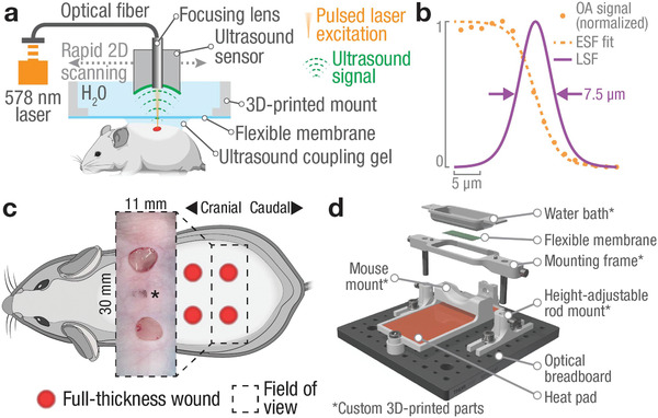
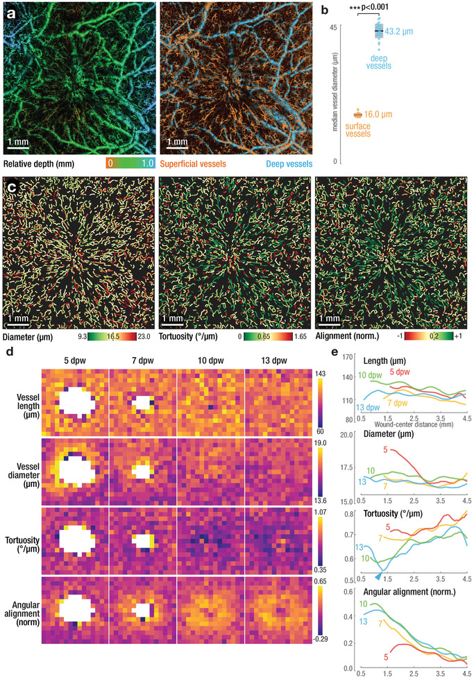
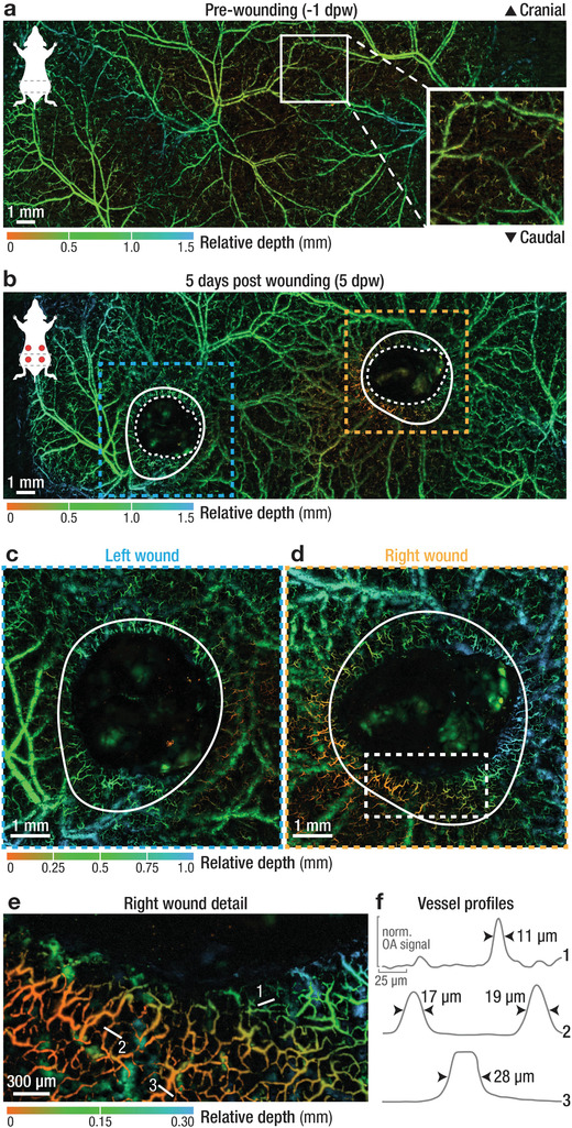
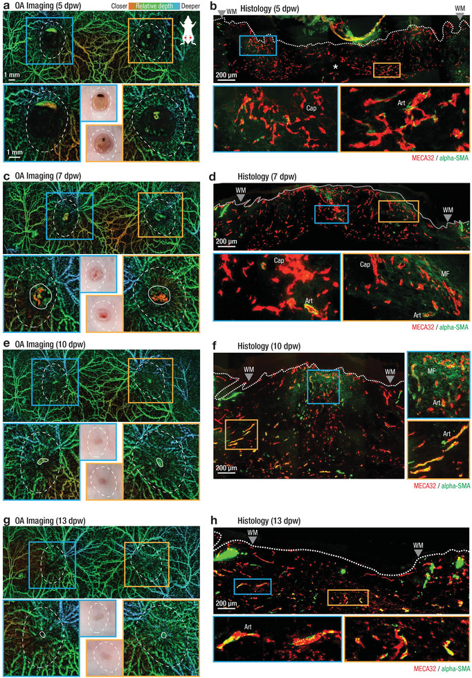
Wound healing is a well-coordinated process, necessitating efficient formation of new blood vessels. Vascularization defects are therefore a major risk factor for chronic, non-healing wounds. The dynamics of mammalian tissue revascularization, vessel maturation, and remodeling remain poorly understood due to lack of suitable in vivo imaging tools. A label-free large-scale optoacoustic microscopy (LSOM) approach is developed for rapid, non-invasive, volumetric imaging of tissue regeneration over large areas spanning up to 50 mm with a depth penetration of 1.5 mm. Vascular networks in dorsal mouse skin and full-thickness excisional wounds are imaged with capillary resolution during the course of healing, revealing previously undocumented views of the angiogenesis process in an unperturbed wound environment. Development of an automatic analysis framework enables the identification of key features of wound angiogenesis, including vessel length, diameter, tortuosity, and angular alignment. The approach offers a versatile tool for preclinical research in tissue engineering and regenerative medicine, empowering label-free, longitudinal, high-throughput, and quantitative studies of the microcirculation in processes associated with normal and impaired vascular remodeling, and analysis of vascular responses to pharmacological interventions in vivo.

摘要

伤口愈合是一个协调良好的过程,需要有效地形成新的血管。因此,血管化缺陷是慢性、不愈合伤口的主要风险因素。由于缺乏合适的体内成像工具,哺乳动物组织再血管化、血管成熟和重塑的动力学仍知之甚少。本研究开发了一种无标记的大规模光声显微镜(LSOM)方法,用于快速、非侵入性、对大区域(最大 50mm)的组织再生进行体积成像,深度穿透可达 1.5mm。在愈合过程中,用毛细血管分辨率对背部小鼠皮肤和全层切除伤口的血管网络进行成像,揭示了在未受干扰的伤口环境中以前未记录的血管生成过程的观点。开发了一种自动分析框架,能够识别伤口血管生成的关键特征,包括血管长度、直径、迂曲度和角向排列。该方法为组织工程和再生医学的临床前研究提供了一种通用工具,能够进行无标记、纵向、高通量和定量的微血管研究,分析正常和受损血管重塑过程中的血管反应,并分析体内药物干预的血管反应。

https://cdn.ncbi.nlm.nih.gov/pmc/blobs/4f01/8261523/7d8598490401/ADVS-8-2004226-g001.jpg
https://cdn.ncbi.nlm.nih.gov/pmc/blobs/4f01/8261523/66ae354e4046/ADVS-8-2004226-g002.jpg
https://cdn.ncbi.nlm.nih.gov/pmc/blobs/4f01/8261523/1a99dabf8b6f/ADVS-8-2004226-g004.jpg
https://cdn.ncbi.nlm.nih.gov/pmc/blobs/4f01/8261523/f91b94fd86c4/ADVS-8-2004226-g003.jpg
https://cdn.ncbi.nlm.nih.gov/pmc/blobs/4f01/8261523/7d8598490401/ADVS-8-2004226-g001.jpg

相似文献

1
Long-Term Imaging of Wound Angiogenesis with Large Scale Optoacoustic Microscopy.大尺度光声显微镜长期成像的伤口血管生成。
Adv Sci (Weinh). 2021 May 2;8(13):2004226. doi: 10.1002/advs.202004226. eCollection 2021 Jul.
2
Non-invasive longitudinal imaging of VEGF-induced microvascular alterations in skin wounds.非侵入性纵向成像技术观察血管内皮生长因子诱导的皮肤创伤中微血管变化。
Theranostics. 2022 Jan 1;12(2):558-573. doi: 10.7150/thno.65287. eCollection 2022.
3
A new model for studying the revascularization of skin grafts in vivo: the role of angiogenesis.一种用于研究体内皮肤移植血管再生的新模型:血管生成的作用。
Plast Reconstr Surg. 2008 Dec;122(6):1669-1680. doi: 10.1097/PRS.0b013e31818cbeb1.
4
Intravital insights in skin wound healing using the mouse dorsal skin fold chamber.利用小鼠背部皮肤褶皱腔对皮肤伤口愈合进行活体观察。
J Anat. 2007 Dec;211(6):810-8. doi: 10.1111/j.1469-7580.2007.00822.x. Epub 2007 Nov 13.
5
Tracking Strain-Specific Morphogenesis and Angiogenesis of Murine Calvaria with Large-Scale Optoacoustic and Ultrasound Microscopy.利用大规模光声和超声显微镜追踪小鼠颅骨的应变特异性形态发生和血管生成。
J Bone Miner Res. 2022 May;37(5):1032-1043. doi: 10.1002/jbmr.4533. Epub 2022 Mar 10.
6
MicroCT angiography detects vascular formation and regression in skin wound healing.显微CT血管造影术可检测皮肤伤口愈合过程中的血管形成与消退。
Microvasc Res. 2016 Jul;106:57-66. doi: 10.1016/j.mvr.2016.03.006. Epub 2016 Mar 21.
7
Real-time analysis of the kinetics of angiogenesis and vascular permeability in an animal model of wound healing.在伤口愈合动物模型中对血管生成和血管通透性动力学进行实时分析。
Burns. 2009 Sep;35(6):811-7. doi: 10.1016/j.burns.2008.12.012. Epub 2009 May 6.
8
Dynamics of angiogenesis during wound healing: a coupled in vivo and in silico study.创伤愈合过程中的血管生成动力学:体内和计算相结合的研究。
Microcirculation. 2011 Apr;18(3):183-97. doi: 10.1111/j.1549-8719.2010.00076.x.
9
[Live Imaging of Angiogenesis during Wound Healing].[伤口愈合过程中血管生成的活体成像]
Yakugaku Zasshi. 2020;140(4):513-519. doi: 10.1248/yakushi.19-00221-2.
10
Optoacoustic mesoscopy for biomedicine.光声介观成像在生物医学中的应用。
Nat Biomed Eng. 2019 May;3(5):354-370. doi: 10.1038/s41551-019-0377-4. Epub 2019 Apr 15.

引用本文的文献

1
Recombinant human collagen type III microgel: an advanced injectable dermal filler for rejuvenating aging skin.重组人III型胶原蛋白微凝胶:一种用于改善老化皮肤的先进可注射真皮填充剂。
Regen Biomater. 2025 Jul 28;12:rbaf076. doi: 10.1093/rb/rbaf076. eCollection 2025.
2
Catalyst-modulated hydrogel dynamics for decoupling viscoelasticity and directing macrophage fate for diabetic wound healing.催化剂调控水凝胶动力学用于解耦粘弹性并引导巨噬细胞命运以促进糖尿病伤口愈合
Bioact Mater. 2025 Jul 5;52:878-895. doi: 10.1016/j.bioactmat.2025.06.007. eCollection 2025 Oct.
3
Local laser fluence estimation in optical resolution optoacoustic angiography employing calibrated ultrasound detector.

本文引用的文献

1
High resolution ultrasound imaging for repeated measure of wound tissue morphometry, biomechanics and hemodynamics under fetal, adult and diabetic conditions.高分辨率超声成像技术用于在胎儿、成人和糖尿病等条件下重复测量伤口组织形态计量学、生物力学和血液动力学。
PLoS One. 2020 Nov 23;15(11):e0241831. doi: 10.1371/journal.pone.0241831. eCollection 2020.
2
Rapid functional optoacoustic micro-angiography in a burst mode.快速功能光声微血管成像的突发模式。
Opt Lett. 2020 May 1;45(9):2522-2525. doi: 10.1364/OL.387630.
3
Intravital optoacoustic and ultrasound bio-microscopy reveal radiation-inhibited skull angiogenesis.
采用校准超声探测器的光学分辨率光声血管造影术中的局部激光能量密度估计
Photoacoustics. 2025 May 29;44:100734. doi: 10.1016/j.pacs.2025.100734. eCollection 2025 Aug.
4
Wound management materials and technologies from bench to bedside and beyond.从实验室到临床乃至更广泛应用的伤口管理材料与技术
Nat Rev Mater. 2024 Aug;9(8):550-566. doi: 10.1038/s41578-024-00693-y. Epub 2024 Jun 17.
5
Characterization of cutaneous microvasculature using 3D imaging: A feasibility study in a cohort of head and neck surgery patients with attention to smoking status.使用三维成像技术对皮肤微血管系统进行特征描述:一项针对头颈外科手术患者队列的可行性研究,重点关注吸烟状况。
JPRAS Open. 2025 Apr 4;44:493-505. doi: 10.1016/j.jpra.2025.03.020. eCollection 2025 Jun.
6
The Potential of Photoacoustic Imaging in Detecting and Managing Complex Wounds.光声成像在复杂伤口检测与处理中的潜力
Biomater Res. 2025 May 21;29:0206. doi: 10.34133/bmr.0206. eCollection 2025.
7
Deletion of microsomal epoxide hydrolase gene leads to increased density in cerebral vasculature and enhances cerebral blood flow in mice.微粒体环氧化物水解酶基因的缺失导致小鼠脑血管密度增加并增强脑血流量。
J Cereb Blood Flow Metab. 2025 Apr 12:271678X251333234. doi: 10.1177/0271678X251333234.
8
Skin layer-specific spatiotemporal assessment of micrometabolism during wound angiogenesis.伤口血管生成过程中微代谢的皮肤层特异性时空评估
Commun Biol. 2024 Dec 20;7(1):1574. doi: 10.1038/s42003-024-07257-4.
9
Enhanced dual-mode imaging: Superior photoacoustic and ultrasound endoscopy in live pigs using a transparent ultrasound transducer.增强双模成像:使用透明超声换能器的活体猪的优越光声和超声内窥镜检查。
Sci Adv. 2024 Nov 22;10(47):eadq9960. doi: 10.1126/sciadv.adq9960.
10
Evaluations of a Cutaneous Wound Healing Model Using Oxygen Enhanced - Dynamic Contrast Enhanced Photoacoustic Imaging (OE-DCE PAI).使用氧增强 - 动态对比增强光声成像(OE-DCE PAI)对皮肤伤口愈合模型的评估
Mol Imaging Biol. 2024 Dec;26(6):995-1004. doi: 10.1007/s11307-024-01966-2. Epub 2024 Nov 12.
活体光声和超声生物显微镜显示辐射抑制颅骨血管生成。
Bone. 2020 Apr;133:115251. doi: 10.1016/j.bone.2020.115251. Epub 2020 Jan 21.
4
Optical-resolution photoacoustic microscopy for monitoring vascular normalization during anti-angiogenic therapy.用于监测抗血管生成治疗期间血管正常化的光学分辨率光声显微镜
Photoacoustics. 2019 Aug 12;15:100143. doi: 10.1016/j.pacs.2019.100143. eCollection 2019 Sep.
5
Review on practical photoacoustic microscopy.实用光声显微镜综述。
Photoacoustics. 2019 Aug 9;15:100141. doi: 10.1016/j.pacs.2019.100141. eCollection 2019 Sep.
6
An in vivo wound healing model for the characterization of the angiogenic process and its modulation by pharmacological interventions.一种用于表征血管生成过程及其通过药物干预进行调节的体内创伤愈合模型。
Sci Rep. 2019 Apr 12;9(1):6004. doi: 10.1038/s41598-019-42479-1.
7
Monitoring neovascularization and integration of decellularized human scaffolds using photoacoustic imaging.使用光声成像监测脱细胞人支架的新生血管形成和整合
Photoacoustics. 2019 Jan 8;13:76-84. doi: 10.1016/j.pacs.2019.01.001. eCollection 2019 Mar.
8
Quantitative analysis on in vivo tumor-microvascular images from optical-resolution photoacoustic microscopy.基于光分辨光声显微镜的活体肿瘤微血管图像的定量分析。
J Biophotonics. 2019 Jun;12(6):e201800421. doi: 10.1002/jbio.201800421. Epub 2019 Mar 13.
9
Methods to label, image, and analyze the complex structural architectures of microvascular networks.标记、成像和分析微血管网络复杂结构体系的方法。
Microcirculation. 2019 Jul;26(5):e12520. doi: 10.1111/micc.12520. Epub 2019 Jan 17.
10
Volumetric Multispectral Optoacoustic Tomography for 3-Dimensional Reconstruction of Skin Tumors: A Further Evaluation with Histopathologic Correlation.用于皮肤肿瘤三维重建的容积多光谱光声断层扫描:与组织病理学相关性的进一步评估
J Invest Dermatol. 2019 Feb;139(2):481-485. doi: 10.1016/j.jid.2018.08.014. Epub 2018 Sep 15.