• 文献检索
  • 文档翻译
  • 深度研究
  • 学术资讯
  • Suppr Zotero 插件Zotero 插件
  • 邀请有礼
  • 套餐&价格
  • 历史记录
应用&插件
Suppr Zotero 插件Zotero 插件浏览器插件Mac 客户端Windows 客户端微信小程序
定价
高级版会员购买积分包购买API积分包
服务
文献检索文档翻译深度研究API 文档MCP 服务
关于我们
关于 Suppr公司介绍联系我们用户协议隐私条款
关注我们

Suppr 超能文献

核心技术专利:CN118964589B侵权必究
粤ICP备2023148730 号-1Suppr @ 2026

文献检索

告别复杂PubMed语法,用中文像聊天一样搜索,搜遍4000万医学文献。AI智能推荐,让科研检索更轻松。

立即免费搜索

文件翻译

保留排版,准确专业,支持PDF/Word/PPT等文件格式,支持 12+语言互译。

免费翻译文档

深度研究

AI帮你快速写综述,25分钟生成高质量综述,智能提取关键信息,辅助科研写作。

立即免费体验

胶原蛋白在人眼筛板和小梁网中的分布。

Collagen distribution in the lamina cribrosa and the trabecular meshwork of the human eye.

作者信息

Rehnberg M, Ammitzböll T, Tengroth B

机构信息

Department of Ophthalmology, Karolinska Institute and Hospital, Stockholm, Sweden.

出版信息

Br J Ophthalmol. 1987 Dec;71(12):886-92. doi: 10.1136/bjo.71.12.886.

DOI:10.1136/bjo.71.12.886
PMID:3426993
原文链接:https://pmc.ncbi.nlm.nih.gov/articles/PMC1041338/
Abstract

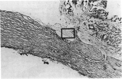
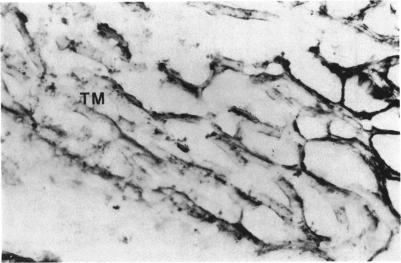
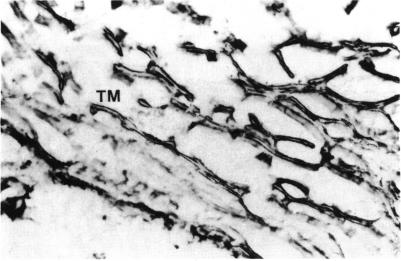
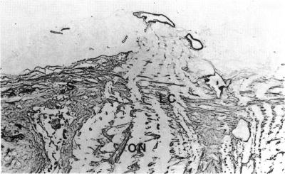
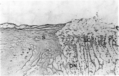
Tengroth and Ammitzböll found the collagen content of the optic disc in glaucoma to differ from that of normal eyes. A theory was advanced that a primary collagen disturbance might be involved in the pathogenesis of glaucoma. The connective tissue in the body has a supportive function in almost all the organs. The tensile strength and elasticity of connective tissue is mainly due to the presence of collagen fibres and elastic fibres, which also maintain the shape of the tissues. There are many different types of collagen, three of which are discussed in this paper. Type I collagen is found in tendons, skin, and numerous other organs, for example the eye. Type III is found mainly in the blood vessels but is also present in other tissues with a mesodermal origin, and type IV is found in the basement membranes. To elucidate the precise distribution of collagen types in the ocular structures an immunhistochemical study was undertaken in normal human eyes. The amino acids proline, hydroxyproline, and hydroxylysine, which are characteristic of collagen, were also analysed. Collagen types I, III, and IV were found in the lamina cribrosa, the trabecular meshwork, and the retrolaminary optic nerve. In contrast, only type I was found in the sclera.

摘要

滕格罗思和阿米茨博尔发现青光眼患者视盘的胶原蛋白含量与正常眼睛不同。有人提出一种理论,认为原发性胶原蛋白紊乱可能参与青光眼的发病机制。身体中的结缔组织在几乎所有器官中都具有支撑功能。结缔组织的抗张强度和弹性主要归因于胶原纤维和弹性纤维的存在,它们也维持着组织的形状。胶原蛋白有许多不同类型,本文讨论其中三种。I型胶原蛋白存在于肌腱、皮肤和许多其他器官中,例如眼睛。III型主要存在于血管中,但也存在于其他中胚层起源的组织中,IV型存在于基底膜中。为了阐明胶原蛋白类型在眼部结构中的精确分布,对正常人眼进行了免疫组织化学研究。还分析了作为胶原蛋白特征的氨基酸脯氨酸、羟脯氨酸和羟赖氨酸。在筛板、小梁网和筛板后视神经中发现了I型、III型和IV型胶原蛋白。相比之下,巩膜中仅发现I型。

https://cdn.ncbi.nlm.nih.gov/pmc/blobs/2d1c/1041338/2b34822c1af2/brjopthal00622-0007-b.jpg
https://cdn.ncbi.nlm.nih.gov/pmc/blobs/2d1c/1041338/225bf1c77d16/brjopthal00622-0004-a.jpg
https://cdn.ncbi.nlm.nih.gov/pmc/blobs/2d1c/1041338/43a4c4cd5770/brjopthal00622-0004-b.jpg
https://cdn.ncbi.nlm.nih.gov/pmc/blobs/2d1c/1041338/5021fc16ae08/brjopthal00622-0005-a.jpg
https://cdn.ncbi.nlm.nih.gov/pmc/blobs/2d1c/1041338/b4e3ae926e2d/brjopthal00622-0006-a.jpg
https://cdn.ncbi.nlm.nih.gov/pmc/blobs/2d1c/1041338/6bff6a555b6e/brjopthal00622-0006-b.jpg
https://cdn.ncbi.nlm.nih.gov/pmc/blobs/2d1c/1041338/325b009296e3/brjopthal00622-0007-a.jpg
https://cdn.ncbi.nlm.nih.gov/pmc/blobs/2d1c/1041338/2b34822c1af2/brjopthal00622-0007-b.jpg

相似文献

1
Collagen distribution in the lamina cribrosa and the trabecular meshwork of the human eye.胶原蛋白在人眼筛板和小梁网中的分布。
Br J Ophthalmol. 1987 Dec;71(12):886-92. doi: 10.1136/bjo.71.12.886.
2
A comparative analysis of the collagen type and distribution in the trabecular meshwork, sclera, lamina cribrosa and the optic nerve in the human eye.人眼小梁网、巩膜、筛板和视神经中胶原蛋白类型及分布的比较分析。
Acta Ophthalmol Suppl (1985). 1985;173:91-3. doi: 10.1111/j.1755-3768.1985.tb06856.x.
3
Changes in the content and composition of collagen in the glaucomatous eye--basis for a new hypothesis for the genesis of chronic open angle glaucoma--a preliminary report.青光眼眼中胶原蛋白含量及成分的变化——慢性开角型青光眼发病新假说的基础——初步报告
Acta Ophthalmol (Copenh). 1984 Dec;62(6):999-1008. doi: 10.1111/j.1755-3768.1984.tb08452.x.
4
Localization of myocilin/trabecular meshwork--inducible glucocorticoid response protein in the human eye.人眼中肌纤蛋白/小梁网诱导性糖皮质激素反应蛋白的定位
Invest Ophthalmol Vis Sci. 2000 Mar;41(3):729-40.
5
Immunohistochemical distribution of type VI collagen in normal and glaucomatous human trabecular meshwork.VI型胶原在正常和青光眼患者人小梁网中的免疫组织化学分布
Graefes Arch Clin Exp Ophthalmol. 1998 Aug;236(8):597-601. doi: 10.1007/s004170050128.
6
Corneal Stiffness and Collagen Cross-Linking Proteins in Glaucoma: Potential for Novel Therapeutic Strategy.青光眼的角膜硬度和胶原交联蛋白:潜在的新型治疗策略。
J Ocul Pharmacol Ther. 2020 Oct;36(8):582-594. doi: 10.1089/jop.2019.0118. Epub 2020 Jul 13.
7
Quantitative analysis of collagen content and amino acids in trabecular meshwork.小梁网中胶原蛋白含量和氨基酸的定量分析。
Br J Ophthalmol. 1990 May;74(5):280-2. doi: 10.1136/bjo.74.5.280.
8
Ultrastructural localization of collagen IV, fibronectin, and laminin in the trabecular meshwork of normal and glaucomatous eyes.正常眼和青光眼眼中小梁网中IV型胶原、纤连蛋白和层粘连蛋白的超微结构定位
Ophthalmic Res. 2001 Nov-Dec;33(6):314-24. doi: 10.1159/000055687.
9
Localization of MYOC transcripts in human eye and optic nerve by in situ hybridization.通过原位杂交对人眼和视神经中MYOC转录本进行定位。
Invest Ophthalmol Vis Sci. 2000 Oct;41(11):3420-8.
10
Distribution of myocilin and extracellular matrix components in the corneoscleral meshwork of human eyes.
Invest Ophthalmol Vis Sci. 2003 Nov;44(11):4772-9. doi: 10.1167/iovs.02-1002.

引用本文的文献

1
Extracellular vesicles as emerging players in glaucoma: Mechanisms, biomarkers, and therapeutic targets.细胞外囊泡在青光眼中成为新角色:机制、生物标志物及治疗靶点
Vision Res. 2025 Jan;226:108522. doi: 10.1016/j.visres.2024.108522. Epub 2024 Nov 23.
2
Pachychoroid pigment epitheliopathy in keratoconic eyes.圆锥角膜眼中的厚脉络膜色素上皮病变
Graefes Arch Clin Exp Ophthalmol. 2025 Jan;263(1):87-95. doi: 10.1007/s00417-024-06612-9. Epub 2024 Aug 30.
3
Characteristic Differences between Normotensive and Hypertensive Pseudoexfoliative Glaucoma.

本文引用的文献

1
Collagen: an overview.胶原蛋白:概述
Methods Enzymol. 1982;82 Pt A:3-32. doi: 10.1016/0076-6879(82)82058-2.
血压正常和高血压性假性剥脱性青光眼之间的特征差异。
J Clin Med. 2024 Feb 14;13(4):1078. doi: 10.3390/jcm13041078.
4
Posterior corneoscleral limbus: Architecture, stem cells, and clinical implications.后角巩膜缘:结构、干细胞及临床意义。
Prog Retin Eye Res. 2023 Sep;96:101192. doi: 10.1016/j.preteyeres.2023.101192. Epub 2023 Jun 29.
5
The effect of ET1-CTGF mediated pathway on the accumulation of extracellular matrix in the trabecular meshwork and its contribution to the increase in IOP.内皮素 1-结缔组织生长因子介导的通路对小梁网细胞外基质积累的影响及其对眼压升高的贡献。
Int Ophthalmol. 2023 Sep;43(9):3297-3307. doi: 10.1007/s10792-023-02733-y. Epub 2023 May 9.
6
Morphological and biomechanical analyses of the human healthy and glaucomatous aqueous outflow pathway: Imaging-to-modeling.人健康和青光眼房水流出通路的形态学和生物力学分析:从成像到建模。
Comput Methods Programs Biomed. 2023 Jun;236:107485. doi: 10.1016/j.cmpb.2023.107485. Epub 2023 Mar 16.
7
Developing an experimental-computational workflow to study the biomechanics of the human conventional aqueous outflow pathway.开发一种实验计算工作流程,以研究人类传统房水流出途径的生物力学。
Acta Biomater. 2023 Jul 1;164:346-362. doi: 10.1016/j.actbio.2023.04.008. Epub 2023 Apr 16.
8
Downregulation of COL12A1 and COL13A1 by a selective EP2 receptor agonist, omidenepag, in human trabecular meshwork cells.选择性 EP2 受体激动剂 omidenepag 下调人眼小梁细胞中 COL12A1 和 COL13A1 的表达。
PLoS One. 2023 Jan 11;18(1):e0280331. doi: 10.1371/journal.pone.0280331. eCollection 2023.
9
Biomechanics of human trabecular meshwork in healthy and glaucoma eyes via dynamic Schlemm's canal pressurization.通过动态施莱姆氏管加压研究健康眼和青光眼眼中的人眼小梁网的生物力学。
Comput Methods Programs Biomed. 2022 Jun;221:106921. doi: 10.1016/j.cmpb.2022.106921. Epub 2022 May 27.
10
Modeling the biomechanics of the conventional aqueous outflow pathway microstructure in the human eye.建立人眼传统房水流出通路微观结构的生物力学模型。
Comput Methods Programs Biomed. 2022 Jun;221:106922. doi: 10.1016/j.cmpb.2022.106922. Epub 2022 May 29.