Bermond Katharina, Berlin Andreas, Tarau Ioana-Sandra, Wobbe Christina, Heintzmann Rainer, Curcio Christine A, Sloan Kenneth R, Ach Thomas
Department of Ophthalmology, University Hospital Würzburg, Würzburg, Germany.
Department of Ophthalmology, Ludwigshafen Hospital, Ludwigshafen, Germany.
Ann Eye Sci. 2021 Mar;6. doi: 10.21037/aes-2021-01. Epub 2021 Mar 15.
Cells of the retinal pigment epithelium (RPE) accumulate different kinds of granules (lipofuscin, melanolipofuscin, melanosomes) within their cell bodies, with lipofuscin and melanolipofuscin being autofluorescent after blue light excitation. High amounts of lipofuscin granules within the RPE have been associated with the development of RPE cell death and age-related macular degeneration (AMD); however, this has not been confirmed in histology so far. Here, based on our previous dataset of RPE granule characteristics, we report the characteristics of RPE cells from human donor eyes that show either high or low numbers of intracellular granules or high or low autofluorescence (AF) intensities.
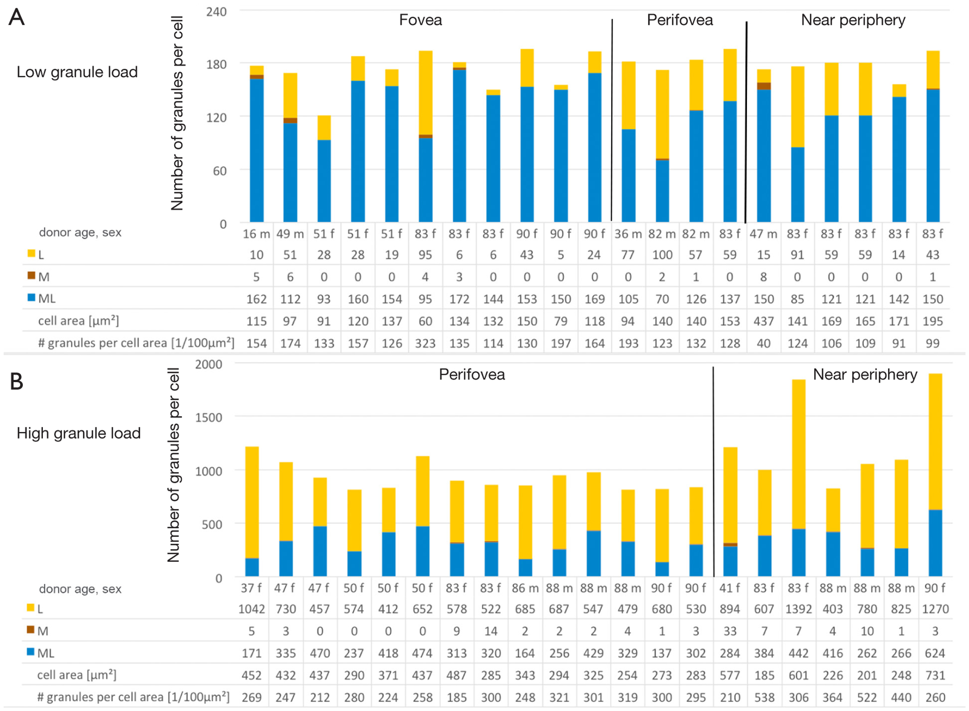
RPE flatmounts of fifteen human donors were examined using high-resolution structured illumination microscopy (HR-SIM) and laser scanning microscopy (LSM). Autofluorescent granules were analyzed regarding AF phenotype and absolute number of granules. In addition, total AF intensity per cell and granule density (number of granules per cell area) were determined. For the final analysis, RPE cells with total granule number below 5 or above the 95 percentile, or a total AF intensity ± 1.5 standard deviations above or below the mean were included, and compared to the average RPE cell at the same location. Data are presented as mean ± standard deviation.
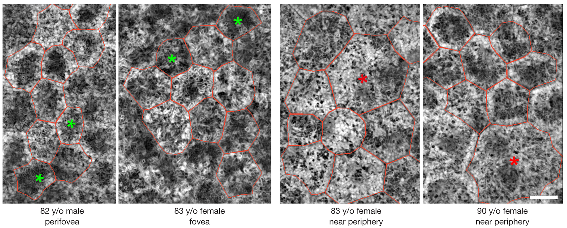
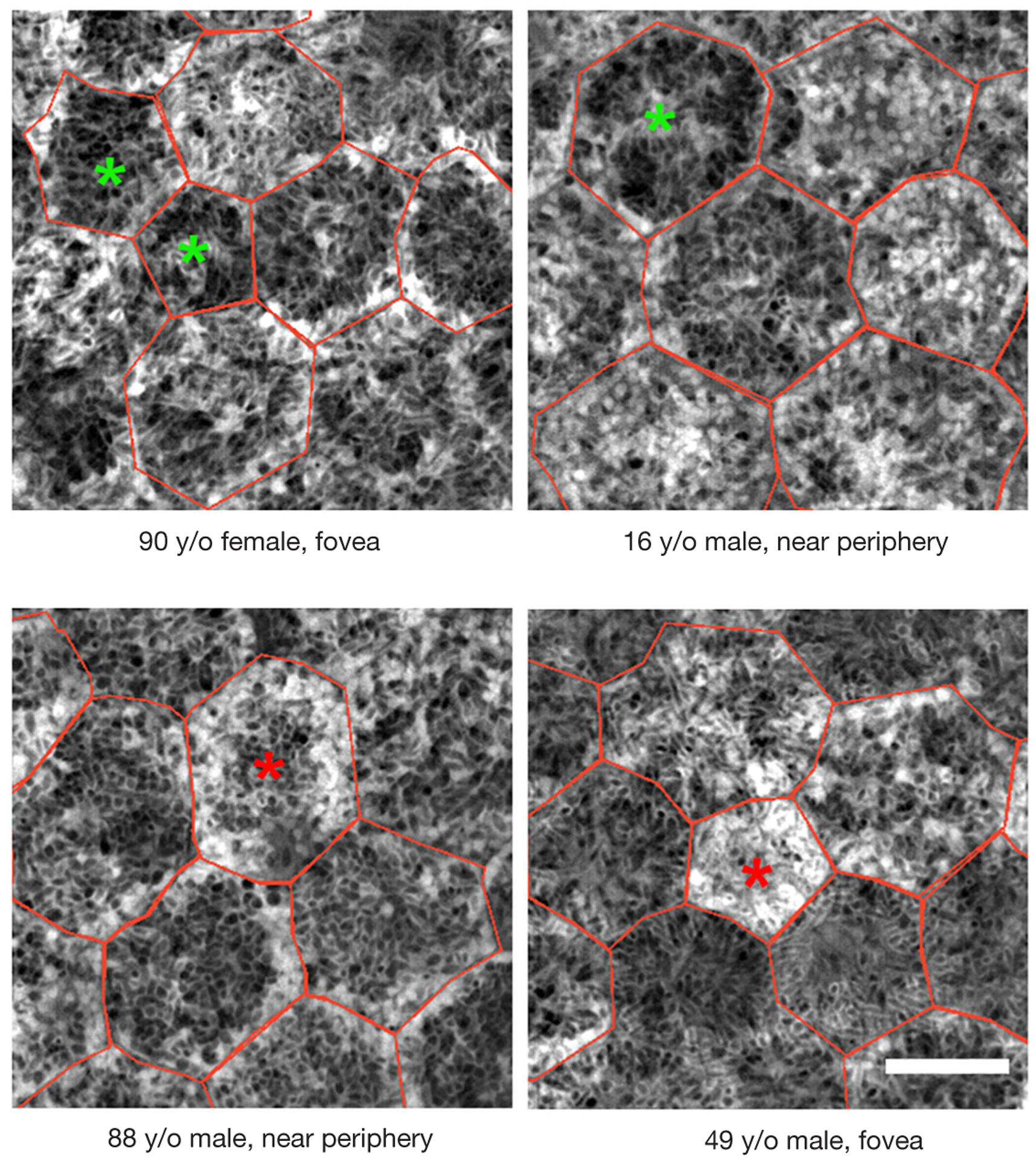
Within 420 RPE cells examined, 42 cells were further analyzed due to extremes regarding total granule numbers. In addition, 20 RPE cells had AF 1.5 standard deviations below, 28 RPE cells above the mean local AF intensity. Melanolipofuscin granules predominate in RPE cells with low granule content and low AF intensity. RPE cells with high granule content have nearly twice (1.8 times) as many granules as an average RPE cell.
In normal eyes, outliers regarding autofluorescent granule load and AF intensity signals are rare among RPE cells, suggesting that granule deposition and subsequent AF follows intrinsic control mechanisms at a cellular level. The AF of a cell is related to the composition of intracellular granule types. Ongoing studies using AMD donor eyes will examine possible disease related changes in granule distribution and further put lipofuscińs role in aging and AMD further into perspective.
视网膜色素上皮(RPE)细胞在其胞体内积累不同种类的颗粒(脂褐素、黑素脂褐素、黑素小体),脂褐素和黑素脂褐素在蓝光激发后会自发荧光。RPE细胞内大量的脂褐素颗粒与RPE细胞死亡及年龄相关性黄斑变性(AMD)的发生有关;然而,到目前为止,这一点在组织学上尚未得到证实。在此,基于我们之前关于RPE颗粒特征的数据集,我们报告了来自人类供体眼的RPE细胞的特征,这些细胞显示出细胞内颗粒数量的高或低,或自发荧光(AF)强度的高或低。
使用高分辨率结构照明显微镜(HR - SIM)和激光扫描显微镜(LSM)检查了15名人类供体的RPE平铺标本。对自发荧光颗粒的AF表型和颗粒绝对数量进行了分析。此外,还测定了每个细胞的总AF强度和颗粒密度(每细胞面积的颗粒数量)。在最终分析中,纳入了总颗粒数低于5或高于第95百分位数,或总AF强度高于或低于平均值±1.5个标准差的RPE细胞,并与同一位置的平均RPE细胞进行比较。数据以平均值±标准差表示。
在所检查的420个RPE细胞中,由于总颗粒数处于极端情况,有42个细胞被进一步分析。此外,有20个RPE细胞的AF低于平均局部AF强度1.5个标准差,28个RPE细胞高于平均局部AF强度。黑素脂褐素颗粒在颗粒含量低和AF强度低的RPE细胞中占主导地位。颗粒含量高的RPE细胞的颗粒数量几乎是平均RPE细胞的两倍(1.8倍)。
在正常眼中,RPE细胞中自发荧光颗粒负荷和AF强度信号的异常值很少见,这表明颗粒沉积及随后的AF在细胞水平上遵循内在控制机制。细胞的AF与细胞内颗粒类型的组成有关。正在使用AMD供体眼进行的研究将检查颗粒分布中可能与疾病相关的变化,并进一步阐明脂褐素在衰老和AMD中的作用。