Le Kang, Sun Jing, Khawaja Hunain, Shibata Maho, Maggirwar Sanjay B, Smith Mitchell R, Gupta Mamta
Department of Biochemistry and Molecular Medicine.
Department of Anatomy and Cell Biology.
Blood Adv. 2021 Jul 27;5(14):2863-2878. doi: 10.1182/bloodadvances.2020003871.
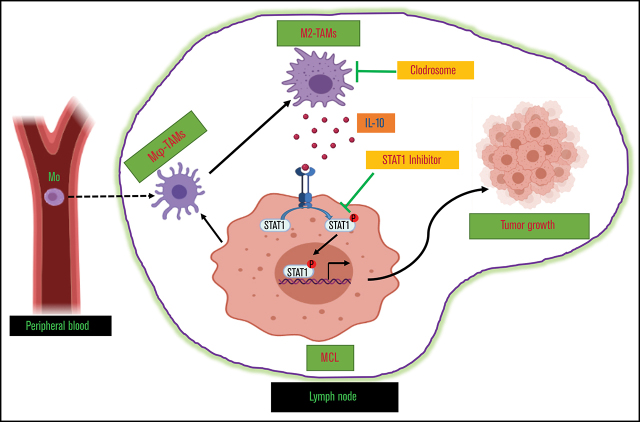
Tumor-associated macrophages (TAMs) are recognized as a hallmark of certain solid cancers and predictors of poor prognosis; however, the functional role of TAMs in lymphoid malignancies, including B-cell lymphoma, has not been well defined. We identified infiltration of F4/80+ TAMs in a syngeneic mouse model using the recently generated murine mantle cell lymphoma (MCL) cell line FC-muMCL1. Multicolor flow cytometric analysis of syngeneic lymphoma tumors showed distinct polarization of F4/80+ TAMs into CD206+ M2 and CD80+ M1 phenotypes. Using human MCL cell lines (Mino, Granta, and JVM2), we further showed that MCL cells polarized monocyte-derived macrophages toward an M2-like phenotype, as assessed by CD163+ expression and increased interleukin-10 (IL-10) level; however, levels of the M1 markers CD80 and IL-12 remained unaffected. To show that macrophages contribute to MCL tumorigenesis, we xenografted the human MCL cell line Mino along with CD14+ monocytes and compared tumor growth between these 2 groups. Results showed that xenografted Mino along with CD14+ monocytes significantly increased the tumor growth in vivo compared with MCL cells alone (P < .001), whereas treatment with liposomal clodronate (to deplete the macrophages) reversed the effect of CD14+ monocytes on growth of MCL xenografts (P < .001). Mechanistically, IL-10 secreted by MCL-polarized M2-like macrophages was found to be responsible for increasing MCL growth by activating STAT1 signaling, whereas IL-10 neutralizing antibody or STAT1 inhibition by fludarabine or STAT1 short hairpin RNA significantly abolished MCL growth (P < .01). Collectively, our data show the existence of a tumor microenvironmental network of macrophages and MCL tumor and suggest the importance of macrophages in interventional therapeutic strategies against MCL and other lymphoid malignancies.
肿瘤相关巨噬细胞(TAM)被认为是某些实体癌的一个标志以及预后不良的预测指标;然而,TAM在包括B细胞淋巴瘤在内的淋巴系统恶性肿瘤中的功能作用尚未明确界定。我们利用最近构建的小鼠套细胞淋巴瘤(MCL)细胞系FC-muMCL1,在同基因小鼠模型中鉴定出F4/80+ TAM的浸润情况。对同基因淋巴瘤肿瘤进行的多色流式细胞术分析显示,F4/80+ TAM明显极化为CD206+ M2和CD80+ M1表型。利用人MCL细胞系(Mino、Granta和JVM2),我们进一步表明,MCL细胞使单核细胞衍生的巨噬细胞极化为M2样表型,这通过CD163+表达和白细胞介素-10(IL-10)水平升高得以评估;然而,M1标志物CD80和IL-12的水平未受影响。为了证明巨噬细胞有助于MCL的肿瘤发生,我们将人MCL细胞系Mino与CD14+单核细胞进行异种移植,并比较这两组之间的肿瘤生长情况。结果显示,与单独的MCL细胞相比,将Mino与CD14+单核细胞一起异种移植显著增加了体内肿瘤生长(P <.001),而用脂质体氯膦酸盐(以清除巨噬细胞)进行治疗逆转了CD14+单核细胞对MCL异种移植物生长的影响(P <.001)。从机制上来说,发现MCL极化的M2样巨噬细胞分泌的IL-10通过激活STAT1信号传导来促进MCL生长,而IL-10中和抗体或氟达拉滨对STAT1的抑制作用或STAT1短发夹RNA显著消除了MCL生长(P <.01)。总体而言,我们的数据表明存在巨噬细胞与MCL肿瘤的肿瘤微环境网络,并提示巨噬细胞在针对MCL和其他淋巴系统恶性肿瘤的介入治疗策略中的重要性。