Alenazy Mohammed S, Al-Nazhan Saad, Mosadomi Hezekiah A
East of Riyadh Dental center-Second Health Cluster, Ministry of Health, Riyadh, Kingdom of Saudi Arabia.
Division of Endodontics, Department of Restorative Dentistry, College of Dentistry, Riyadh Elm University, Riyadh, Kingdom of Saudi Arabia.
Curr Ther Res Clin Exp. 2020 Dec 16;94:100620. doi: 10.1016/j.curtheres.2020.100620. eCollection 2021.
Blood clot (BC) and platelet-rich fibrin (PRF) has been successfully used to biologically treat immature roots. It is nowadays considered the treatment of choice.
This study aimed to determine the ability of PRF and BC scaffolds to enhance regeneration of disinfected root canals and healing of apical periodontitis within experimentally enlarged canal apices of dog teeth.
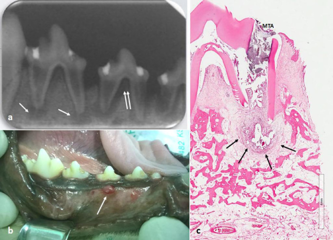
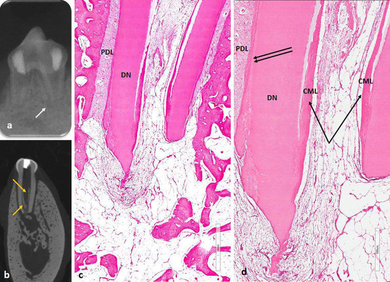
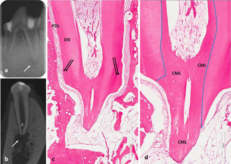
Forty-eight root canals in 28 mandibular premolars from 4 healthy adult dogs were experimentally infected and developed apical periodontitis. The teeth were randomly divided into a control (untreated) group, a disinfection only group, a group that received disinfection and a BC scaffold, and a group that received disinfection and a BC + PRF scaffold. Healing of the apical radiolucency was evaluated by conventional radiography, micro-computed tomography, and histology after 3 months. The data were analyzed by χ test.
Healing was achieved in 49% of roots as seen on radiograph and 43% as seen on micro-computed tomography. There was no significant between-group difference in the presence or absence of periapical radiolucency in the mesial roots when seen on conventional images ( = 0.255), but there was a significant difference in the distal roots ( = 0.001); similarly, on micro-computed tomography, there was no significant between-group difference in the mesial roots ( = 0.174) but there was a significant difference in the distal roots ( = 0.001). Histologically, apical closure was significantly not greater in the BC + PRF scaffold group than in the BC scaffold group ( = 0.001).
A mix of BC + PRF scaffold did not improve tissue regeneration in experimentally enlarged dog teeth. ( 2021; 82:XXX-XXX) © 2021 Elsevier HS Journals, Inc.
血凝块(BC)和富血小板纤维蛋白(PRF)已成功用于未成熟牙根的生物治疗。如今它被视为首选治疗方法。
本研究旨在确定PRF和BC支架增强消毒根管再生以及愈合犬齿实验性扩大根管根尖处根尖周炎的能力。
对4只健康成年犬的28颗下颌前磨牙的48个根管进行实验性感染并引发根尖周炎。将牙齿随机分为对照组(未治疗)、仅消毒组、接受消毒并植入BC支架组以及接受消毒并植入BC + PRF支架组。3个月后,通过传统X线摄影、显微计算机断层扫描和组织学评估根尖透射区的愈合情况。数据采用χ检验进行分析。
X线片显示49%的牙根实现愈合,显微计算机断层扫描显示43%的牙根实现愈合。在传统图像上观察时,近中牙根根尖透射区有无在组间无显著差异(χ² = 0.255),但远中牙根有显著差异(χ² = 0.001);同样,在显微计算机断层扫描上,近中牙根在组间无显著差异(χ² = 0.174),但远中牙根有显著差异(χ² = 0.001)。组织学上,BC + PRF支架组的根尖封闭显著不比BC支架组更大(χ² = 0.001)。
BC + PRF支架组合并未改善实验性扩大犬齿中的组织再生。(2021;82:XXX - XXX)©2021爱思唯尔HS期刊公司