Institute of Pharmacy, Pharmacology and Toxicology, Center for Chemistry and Biomedicine, University of Innsbruck, Innrain 80-82, 6020, Innsbruck, Austria.
CNR Neuroscience Institute, 56124, Pisa, Italy.
Sci Rep. 2021 Jul 26;11(1):15146. doi: 10.1038/s41598-021-94304-3.
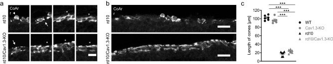
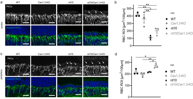
Retinitis Pigmentosa is a genetically heterogeneous, degenerative retinal disorder characterized by gradual dysfunction and death of photoreceptors, first rods and later cones, and progressive blindness. Studies suggested that application of L-type calcium channel blockers rescues photoreceptors in paradigms related to Ca overflow. To investigate whether Cav1.3 L-type channels have protective effects in the retina, we established a new mouse model by crossing rd10, modeling autosomal-recessive RP, with Cav1.3 deficient mice (rd10/Cav1.3KO). Our immunohistochemical analyses revealed an influence of Cav1.3 channels on the degenerative process of photoreceptors. The absence of Cav1.3 delayed the centre-to-periphery degeneration of rods indicated by a significantly higher number of photoreceptor rows and, consequently, of cones. In accordance with a preserved number of cones we observed a regular row of cone somas in rd10/Cav1.3-KO retinas. Surviving rod photoreceptors maintained synaptic contacts with rod bipolar cells. However, the delay in degeneration was only observed up to postnatal day 45. Although we observed a reduction in the spontaneous oscillatory retinal activity during multielectrode array analyses, measurable functional preservation was lacking in behavioural tests. In conclusion, Cav1.3 channels contribute to photoreceptor degeneration in rd10 retinas but photoreceptor temporary rescue might rather be achieved indirectly through other retinal cell layers.
色素性视网膜炎是一种遗传性、退行性视网膜疾病,其特征是感光细胞(首先是视杆细胞,然后是视锥细胞)逐渐功能障碍和死亡,并导致进行性失明。研究表明,L 型钙通道阻滞剂的应用可挽救与 Ca 溢出相关的模型中的感光细胞。为了研究 Cav1.3 L 型通道是否对视网膜具有保护作用,我们通过将 rd10(模拟常染色体隐性 RP)与 Cav1.3 缺陷小鼠(rd10/Cav1.3KO)杂交,建立了一种新的小鼠模型。我们的免疫组织化学分析表明 Cav1.3 通道对感光细胞的退行性过程有影响。Cav1.3 的缺失延迟了 rods 的中心到周边的退化,这表现为感光细胞行数(以及随后的 cone 行数)显著增加。与 cone 数量保持一致,我们观察到 rd10/Cav1.3-KO 视网膜中的 cone 体呈规则的一行排列。存活的 rod 感光细胞保持与 rod 双极细胞的突触接触。然而,这种退化延迟仅在出生后 45 天内观察到。尽管我们在多电极阵列分析中观察到自发振荡视网膜活动减少,但行为测试中缺乏可测量的功能保留。总之,Cav1.3 通道有助于 rd10 视网膜中的感光细胞退化,但感光细胞的暂时挽救可能主要是通过其他视网膜细胞层间接实现的。