• 文献检索
  • 文档翻译
  • 深度研究
  • 学术资讯
  • Suppr Zotero 插件Zotero 插件
  • 邀请有礼
  • 套餐&价格
  • 历史记录
应用&插件
Suppr Zotero 插件Zotero 插件浏览器插件Mac 客户端Windows 客户端微信小程序
定价
高级版会员购买积分包购买API积分包
服务
文献检索文档翻译深度研究API 文档MCP 服务
关于我们
关于 Suppr公司介绍联系我们用户协议隐私条款
关注我们

Suppr 超能文献

核心技术专利:CN118964589B侵权必究
粤ICP备2023148730 号-1Suppr @ 2026

文献检索

告别复杂PubMed语法,用中文像聊天一样搜索,搜遍4000万医学文献。AI智能推荐,让科研检索更轻松。

立即免费搜索

文件翻译

保留排版,准确专业,支持PDF/Word/PPT等文件格式,支持 12+语言互译。

免费翻译文档

深度研究

AI帮你快速写综述,25分钟生成高质量综述,智能提取关键信息,辅助科研写作。

立即免费体验

超声评估猫在献血过程中腔静脉直径。

Ultrasonographic assessment of the caudal vena cava diameter in cats during blood donation.

机构信息

Clinic of Small Animal Medicine, Centre for Clinical Veterinary Medicine, Faculty of Veterinary Medicine, Ludwig-Maximilians-University, Munich, Germany.

出版信息

J Feline Med Surg. 2022 Jun;24(6):484-492. doi: 10.1177/1098612X211028838. Epub 2021 Jul 27.

DOI:10.1177/1098612X211028838
PMID:34313488
原文链接:https://pmc.ncbi.nlm.nih.gov/articles/PMC9160942/
Abstract

OBJECTIVES

Ultrasonography of the caudal vena cava (CVC) has been previously established to assess fluid status in dogs but not in cats. The aim of this study was to determine CVC diameter changes during feline blood donation.

METHODS

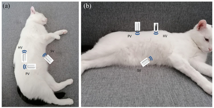
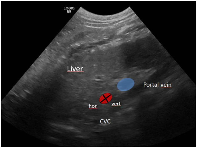
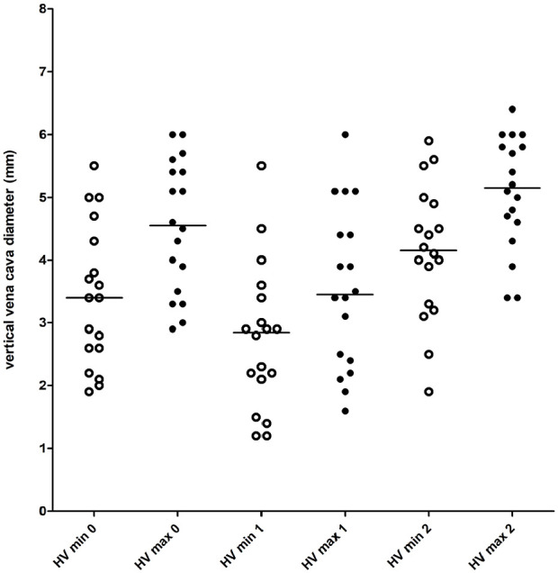
Inter- and intra-observer variability were assessed in 11 client-owned cats. Minimal and maximal CVC diameters were assessed longitudinally in the subxiphoid view (SV) and right paralumbar view (PV), and transversely in the right hepatic intercostal view (HV). Eighteen client-owned, healthy, anaesthetised cats were evaluated during 21 blood donation procedures of 10 ml/kg in the same anatomical locations before (T0) and after (T1) blood donation, and after volume resuscitation with 30 ml/kg lactated Ringer's solution (T2). The CVC index was calculated.

RESULTS

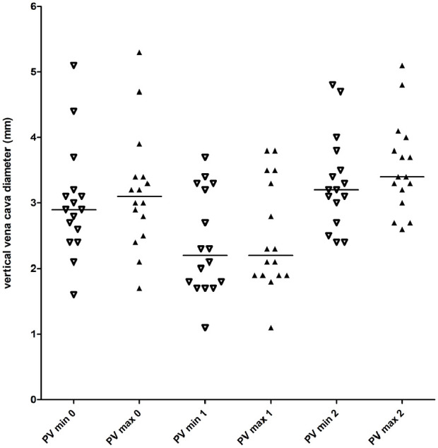
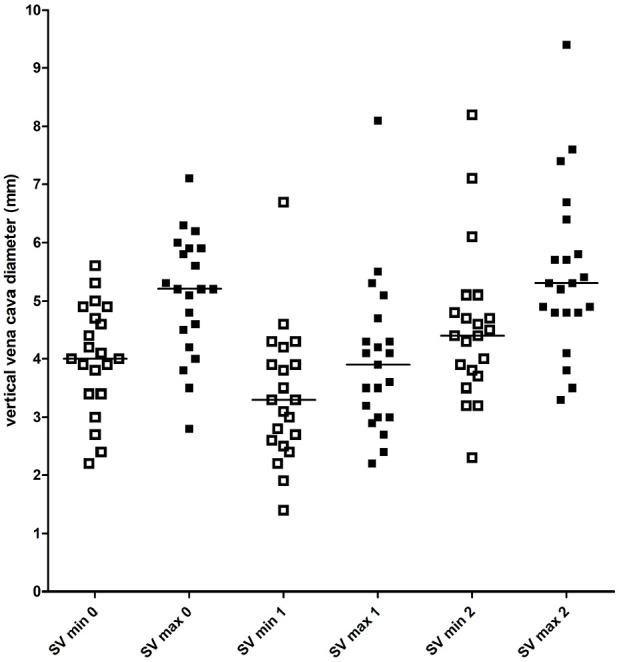
Intra-observer variability was acceptable for all probe positions, except for the HV, whereas inter-observer variability was considered unacceptable for all probe positions. Complete measurements were obtained during 21 blood donations at T0, T1 and T2 at the SV, during 18/21 blood donations at the HV and during 16/21 blood donations at the PV. At the SV, the minimal CVC diameter between T1 and T2 ( <0.001), and the maximal CVC diameter between T0 and T1 and between T1 and T2 ( <0.001) were significantly different. At the HV, the minimal vertical diameter, maximal vertical diameter and minimal horizontal diameter were different between all timepoints ( <0.001). The maximal horizontal diameter was different between T1 and T2 ( = 0.002). At the PV, both diameters were different between all timepoints ( <0.001). The CVC index was not different between timepoints.

CONCLUSION AND RELEVANCE

Significant probe position dependent CVC diameter changes with marked overlap were observed before and after blood donation, and after fluid bolus. No absolute CVC diameter could be used to indicate hypovolaemia. Ultrasonographic assessment of the feline CVC is highly operator-dependent. The CVC index is not useful in cats.

摘要

目的

已经证实,通过对犬尾腔静脉(CVC)进行超声检查可以评估其液体状态,但尚未在猫中进行研究。本研究旨在确定猫在献血过程中 CVC 直径的变化。

方法

在 11 只患有疾病的猫中评估了观察者内和观察者间的变异性。在剑突下(SV)和右肋旁(PV)矢状面以及右肝肋间(HV)横切面纵向评估最小和最大 CVC 直径。在 21 次采血过程中,对 18 只接受麻醉的健康、患有疾病的猫进行了评估,在相同的解剖位置采血 10ml/kg,分别在采血前(T0)、采血后(T1)以及体积复苏后(T2,给予 30ml/kg 乳酸林格氏液)进行评估。计算 CVC 指数。

结果

除 HV 外,所有探头位置的观察者内变异性均在可接受范围内,而所有探头位置的观察者间变异性均被认为不可接受。在 SV 上,在 T0、T1 和 T2 时,在 21 次采血过程中可以获得完整的测量值;在 HV 上,在 18/21 次采血过程中可以获得完整的测量值;在 PV 上,在 16/21 次采血过程中可以获得完整的测量值。在 SV 上,T1 与 T2 之间的最小 CVC 直径(<0.001)和 T0 与 T1 之间以及 T1 与 T2 之间的最大 CVC 直径(<0.001)有显著差异。在 HV 上,最小垂直直径、最大垂直直径和最小水平直径在所有时间点均有差异(<0.001)。T1 与 T2 之间最大水平直径有差异(=0.002)。在 PV 上,两个直径在所有时间点均有差异(<0.001)。CVC 指数在各时间点无差异。

结论

在献血前后以及液体冲击后,观察到明显的与探头位置相关的 CVC 直径变化,并且重叠度较大。没有绝对的 CVC 直径可以用于指示低血容量。猫的 CVC 超声评估高度依赖操作者。CVC 指数在猫中没有用处。

https://cdn.ncbi.nlm.nih.gov/pmc/blobs/a5b2/11104243/521d94c691c3/10.1177_1098612X211028838-fig5.jpg
https://cdn.ncbi.nlm.nih.gov/pmc/blobs/a5b2/11104243/2c31bcc25fb3/10.1177_1098612X211028838-fig1.jpg
https://cdn.ncbi.nlm.nih.gov/pmc/blobs/a5b2/11104243/b7f1c26ad705/10.1177_1098612X211028838-fig2.jpg
https://cdn.ncbi.nlm.nih.gov/pmc/blobs/a5b2/11104243/b05fa6b720be/10.1177_1098612X211028838-fig3.jpg
https://cdn.ncbi.nlm.nih.gov/pmc/blobs/a5b2/11104243/a4d5d1c6e2e4/10.1177_1098612X211028838-fig4.jpg
https://cdn.ncbi.nlm.nih.gov/pmc/blobs/a5b2/11104243/521d94c691c3/10.1177_1098612X211028838-fig5.jpg

相似文献

1
Ultrasonographic assessment of the caudal vena cava diameter in cats during blood donation.超声评估猫在献血过程中腔静脉直径。
J Feline Med Surg. 2022 Jun;24(6):484-492. doi: 10.1177/1098612X211028838. Epub 2021 Jul 27.
2
Establishment of reference values of the caudal vena cava by fast-ultrasonography through different views in healthy dogs.通过快速超声检查在健康犬不同视图下建立尾静脉腔的参考值。
J Vet Intern Med. 2018 Jul;32(4):1308-1318. doi: 10.1111/jvim.15136. Epub 2018 May 10.
3
Ultrasonographically derived caudal vena cava parameters acquired in a standing position and lateral recumbency in healthy, lightly sedated cats: a pilot study.超声获取的健康、轻度镇静猫站立位和侧卧位尾腔静脉参数:一项初步研究。
J Feline Med Surg. 2022 Oct;24(10):1039-1045. doi: 10.1177/1098612X211064697. Epub 2021 Dec 14.
4
Echocardiographic caudal vena cava measurements in healthy cats and in cats with congestive heart failure and non-cardiac causes of cavitary effusions.超声心动图评估健康猫与充血性心力衰竭猫及非心源性胸腹腔积液猫的尾静脉腔测量值。
J Vet Cardiol. 2023 Aug;48:7-18. doi: 10.1016/j.jvc.2023.05.004. Epub 2023 May 10.
5
Interobserver agreement between non-cardiologist veterinarians and a cardiologist after a 6-hour training course for echographic evaluation of basic echocardiographic parameters and caudal vena cava diameter in 15 healthy Beagles.在对15只健康比格犬进行基本超声心动图参数和尾腔静脉直径的超声评估的6小时培训课程后,非心脏病专家兽医与心脏病专家之间的观察者间一致性。
J Vet Emerg Crit Care (San Antonio). 2019 Sep;29(5):495-504. doi: 10.1111/vec.12883. Epub 2019 Aug 27.
6
Ultrasound measurements of the caudal vena cava before and after blood donation in 9 greyhound dogs.对9只灵缇犬献血前后的后腔静脉进行超声测量。
Can Vet J. 2018 Sep;59(9):973-980.
7
Sonographic assessment of volaemia: development and validation of a new method in dogs.犬类血容量的超声评估:一种新方法的开发与验证
J Small Anim Pract. 2018 Mar;59(3):174-182. doi: 10.1111/jsap.12759. Epub 2017 Sep 27.
8
Influence of the Respiratory Cycle on Caudal Vena Cava Diameter Measured by Sonography in Healthy Foals: A Pilot Study.呼吸周期对健康幼驹超声测量的尾腔静脉直径的影响:一项初步研究。
J Vet Intern Med. 2017 Sep;31(5):1556-1562. doi: 10.1111/jvim.14793. Epub 2017 Aug 1.
9
Ultrasonographical Assessment of Caudal Vena Cava Size through Different Views in Healthy Calves: A Pilot Study.通过不同视角对健康犊牛尾腔静脉大小进行超声评估:一项初步研究
Vet Sci. 2022 Jun 22;9(7):308. doi: 10.3390/vetsci9070308.
10
Establishment of Reference Intervals for Caudal Vena Cava-to-Aorta Ratio Measured Ultrasonographically in Healthy Nonsedated Dogs.健康非镇静犬超声测量尾腔静脉-主动脉比值参考区间的建立。
Top Companion Anim Med. 2023 Sep-Dec;56-57:100822. doi: 10.1016/j.tcam.2023.100822. Epub 2023 Oct 5.

引用本文的文献

1
The ultrasonographic evaluation of caudal vena cava diameter before and after fluid replacement in neonatal dehydrated calves with diarrhea.腹泻致脱水新生犊牛补液前后尾腔静脉直径的超声评估
BMC Vet Res. 2025 Jul 2;21(1):424. doi: 10.1186/s12917-025-04759-z.
2
Feline friendly POCUS: how to implement it into your daily practice.猫友好型 POCT:如何将其融入日常实践。
J Feline Med Surg. 2024 Sep;26(9):1098612X241276916. doi: 10.1177/1098612X241276916.
3
Ultrasonographical Assessment of Caudal Vena Cava Size through Different Views in Healthy Calves: A Pilot Study.

本文引用的文献

1
Caval-aortic ratio and caudal vena cava diameter in dogs before and after blood donation.犬献血前后的腔静脉-主动脉比率及尾腔静脉直径
J Vet Emerg Crit Care (San Antonio). 2019 Nov;29(6):643-646. doi: 10.1111/vec.12900. Epub 2019 Oct 18.
2
Interobserver agreement between non-cardiologist veterinarians and a cardiologist after a 6-hour training course for echographic evaluation of basic echocardiographic parameters and caudal vena cava diameter in 15 healthy Beagles.在对15只健康比格犬进行基本超声心动图参数和尾腔静脉直径的超声评估的6小时培训课程后,非心脏病专家兽医与心脏病专家之间的观察者间一致性。
J Vet Emerg Crit Care (San Antonio). 2019 Sep;29(5):495-504. doi: 10.1111/vec.12883. Epub 2019 Aug 27.
3
通过不同视角对健康犊牛尾腔静脉大小进行超声评估:一项初步研究
Vet Sci. 2022 Jun 22;9(7):308. doi: 10.3390/vetsci9070308.
4
Ultrasonographically derived caudal vena cava parameters acquired in a standing position and lateral recumbency in healthy, lightly sedated cats: a pilot study.超声获取的健康、轻度镇静猫站立位和侧卧位尾腔静脉参数:一项初步研究。
J Feline Med Surg. 2022 Oct;24(10):1039-1045. doi: 10.1177/1098612X211064697. Epub 2021 Dec 14.
Inferior vena cava, abdominal aorta, and IVC-to-aorta ratio in healthy Caucasian children: Ultrasound Z-scores according to BSA and age.
健康白种儿童下腔静脉、腹主动脉和 IVC-主动脉比值:BSA 和年龄的超声 Z 评分。
J Cardiol. 2019 Oct;74(4):388-393. doi: 10.1016/j.jjcc.2019.02.021. Epub 2019 Apr 2.
4
Establishment of reference values of the caudal vena cava by fast-ultrasonography through different views in healthy dogs.通过快速超声检查在健康犬不同视图下建立尾静脉腔的参考值。
J Vet Intern Med. 2018 Jul;32(4):1308-1318. doi: 10.1111/jvim.15136. Epub 2018 May 10.
5
Ultrasonographic measurement of caudal vena cava to aorta ratios for determination of volume depletion in normal beagle dogs.通过超声测量正常比格犬尾腔静脉与主动脉的比例以确定血容量不足。
Vet Radiol Ultrasound. 2018 Mar;59(2):203-211. doi: 10.1111/vru.12566. Epub 2017 Oct 10.
6
Sonographic assessment of volaemia: development and validation of a new method in dogs.犬类血容量的超声评估:一种新方法的开发与验证
J Small Anim Pract. 2018 Mar;59(3):174-182. doi: 10.1111/jsap.12759. Epub 2017 Sep 27.
7
Influence of the Respiratory Cycle on Caudal Vena Cava Diameter Measured by Sonography in Healthy Foals: A Pilot Study.呼吸周期对健康幼驹超声测量的尾腔静脉直径的影响:一项初步研究。
J Vet Intern Med. 2017 Sep;31(5):1556-1562. doi: 10.1111/jvim.14793. Epub 2017 Aug 1.
8
Correlation of IVC Diameter and Collapsibility Index With Central Venous Pressure in the Assessment of Intravascular Volume in Critically Ill Patients.在危重症患者血管内容量评估中,下腔静脉直径和塌陷指数与中心静脉压的相关性
Cureus. 2017 Feb 12;9(2):e1025. doi: 10.7759/cureus.1025.
9
The efficacy of sonographic measurement of inferior vena cava diameter as an estimate of central venous pressure.超声测量下腔静脉直径作为中心静脉压评估指标的有效性。
Cardiovasc Ultrasound. 2016 Aug 20;14(1):33. doi: 10.1186/s12947-016-0076-1.
10
Correlation of the ratio of caudal vena cava diameter and aorta diameter with systolic pressure variation in anesthetized dogs.麻醉犬尾腔静脉直径与主动脉直径之比与收缩压变化的相关性
Am J Vet Res. 2016 Feb;77(2):137-43. doi: 10.2460/ajvr.77.2.137.