Zou Yao, Bhat Owais M, Yuan Xinxu, Li Guangbi, Huang Dandan, Guo Yi, Zhou Dan, Li Pin-Lan
Research Center of Experimental Acupuncture Science, Tianjin University of Traditional Chinese Medicine, Tianjin, People's Republic of China.
Department of Pharmacology and Toxicology, Virginia Commonwealth University, School of Medicine, Richmond, VA, USA.
J Inflamm Res. 2021 Jul 24;14:3501-3521. doi: 10.2147/JIR.S312385. eCollection 2021.
Exosomes have been reported to mediate activation of the inflammatory response by secretion of inflammasome products such as IL-1β or IL-18 and that changes in exosomes production or secretion may be a therapeutic target for treatment of a variety of different chronic diseases. The present study tested the hypothesis that exosome-mediated release of NLRP3 inflammasome products instigates the inflammatory response in the lung during emphysema, a type of chronic obstructive pulmonary disease (COPD) and that electroacupuncture (EA) may attenuate emphysema by inhibition of NLRP3 inflammasome activation and consequent inflammation.
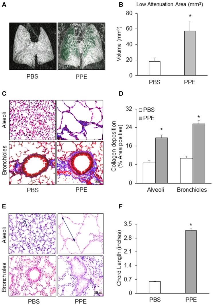
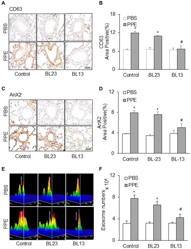
The COPD mice model was developed by injecting porcine pancreatic elastase (PPE) via puncture tracheotomy and instillation. EA (4 Hz/20 Hz, 1 to 3 mA) was applied to the bilateral BL13 and ST36 for 30 min, once every other day for 2 weeks. Micro computed tomography (micro-CT) was performed to measure lung function. Histopathological changes in the lungs were displayed by HE staining.
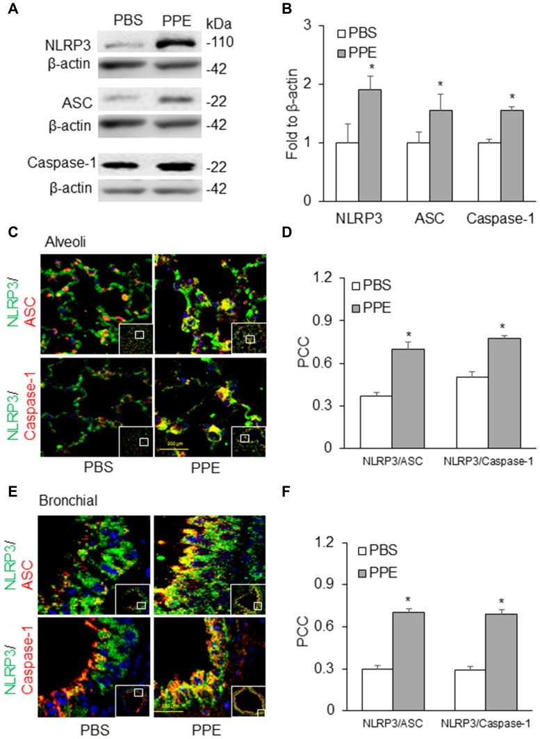
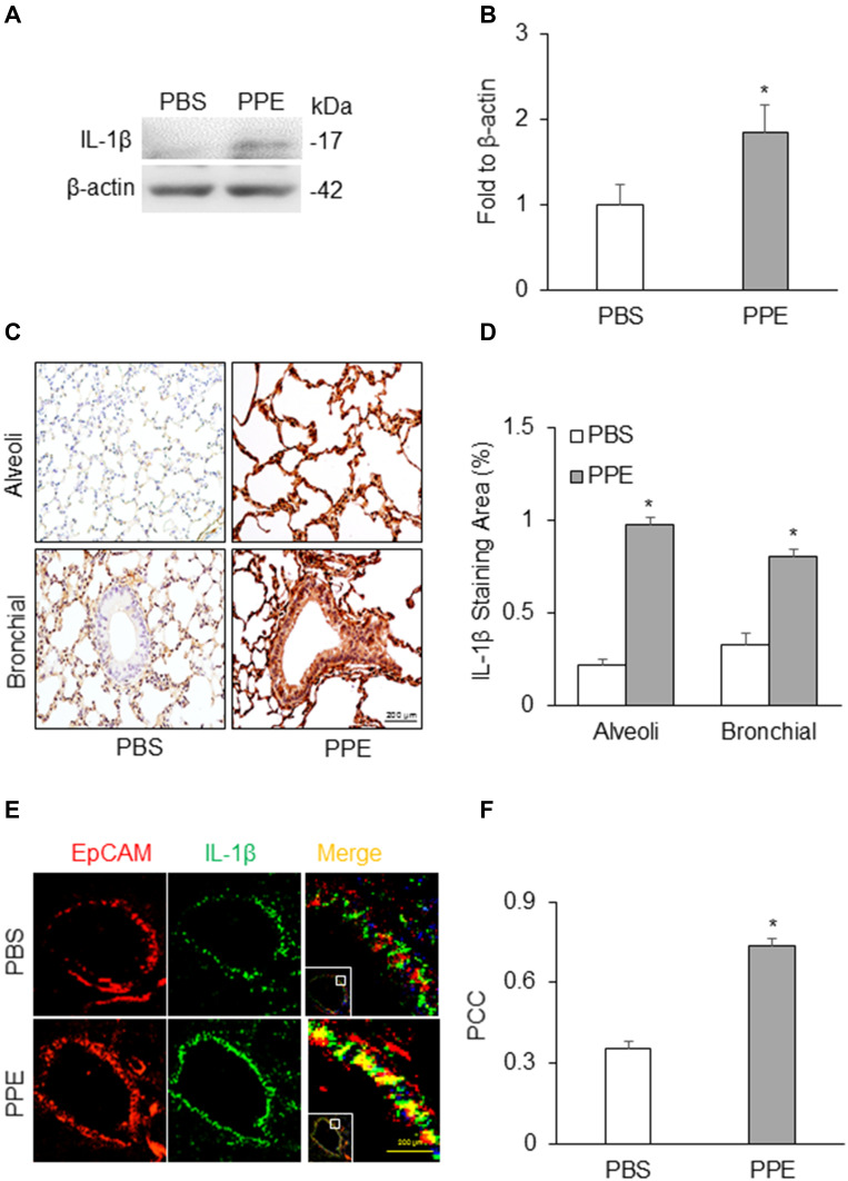
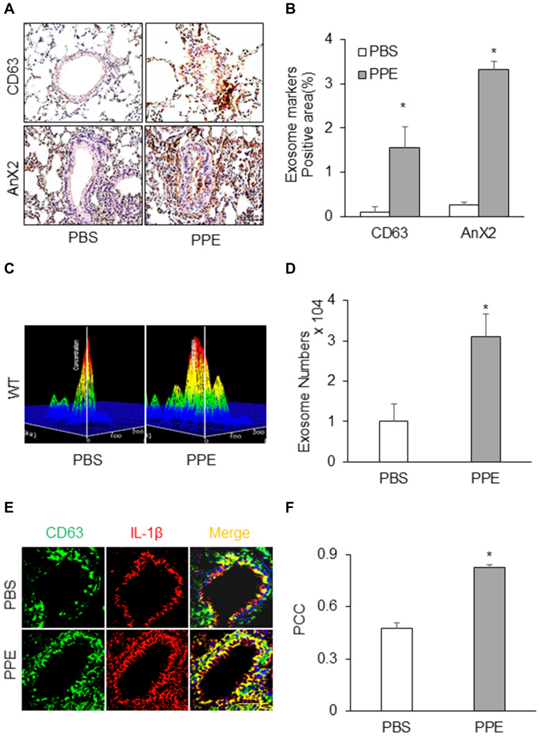
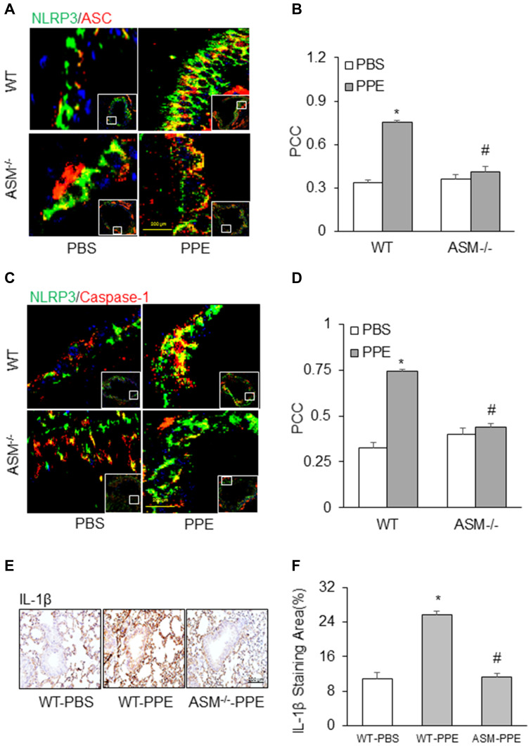
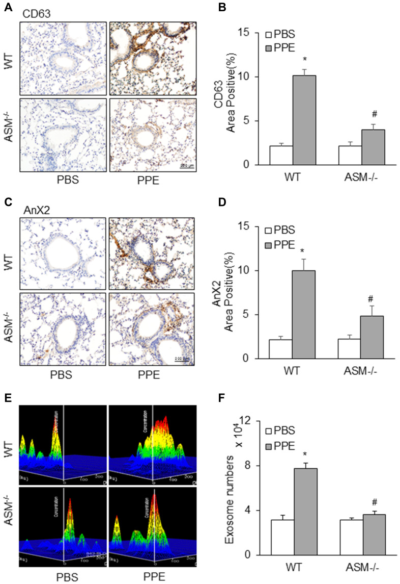
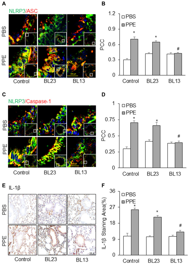
In a mouse model of porcine pancreatic elastase (PPE)-induced emphysema, the lung tissue was found to display several key features of emphysema, including alveolar septal thickening, enlarged alveoli, interstitial edema, and inflammatory cells infiltration. Lungs of mice receiving PPE exhibited substantially increased low attenuation area (LAA) in micro-CT images. The colocalization of NLRP3 vs ASC or caspase-1 detected by confocal microscopy was shown to increase in both bronchial and alveolar walls, indicating the increased formation of NLRP3 inflammasomes. IL-1β, a prototype NLRP3 inflammasome activating product, was also found to have increased in the lung during emphysema, which was colocalized with CD63 (an exosome marker), an indicative of inflammatory exosome formation. By nanoparticle tracking analysis (NTA), IL-1β-containing exosomes were shown to significantly increase in the bronchoalveolar lavage (BAL) from mice with emphysema. Therapeutically, IL-1β production in the lung during emphysema was significantly reduced by EA at the acupoint Feishu (BL13) and Zusanli (ST36), accompanied by decreased colocalization of NLRP3 vs ASC or caspase-1. Increased exosome release into BAL during emphysema was shown to be significantly attenuated in EA-treated mice compared to their controls. However, EA of non-specific BL23 together with ST36 acupoint had no effects on NLRP3 inflammasome activation, exosome release and associated lung pathology during emphysema.
NLRP3 inflammasome activation in concert with increased release of exosomes containing IL-1β or other inflammasome products contributes to the development of lung inflammation and injury during PPE-induced emphysema and that EA of lung-specific acupoints attenuates inflammasome activation and exosome release, thereby reducing inflammatory response in the lung of mice with emphysema.
据报道,外泌体可通过分泌炎性小体产物如白细胞介素-1β(IL-1β)或白细胞介素-18(IL-18)来介导炎症反应的激活,并且外泌体产生或分泌的变化可能是治疗多种不同慢性疾病的治疗靶点。本研究检验了以下假设:外泌体介导的NLRP3炎性小体产物释放引发肺气肿(一种慢性阻塞性肺疾病(COPD))时肺内的炎症反应,并且电针(EA)可能通过抑制NLRP3炎性小体激活及随之而来的炎症来减轻肺气肿。
通过经气管切开穿刺和滴注注射猪胰弹性蛋白酶(PPE)建立COPD小鼠模型。将电针(4Hz/20Hz,1至3mA)施加于双侧肺俞穴(BL13)和足三里穴(ST36)30分钟,每隔一天一次,共2周。进行微型计算机断层扫描(micro-CT)以测量肺功能。通过苏木精-伊红(HE)染色显示肺组织的组织病理学变化。
在猪胰弹性蛋白酶(PPE)诱导的肺气肿小鼠模型中,发现肺组织呈现肺气肿的几个关键特征,包括肺泡间隔增厚、肺泡扩大、间质水肿和炎性细胞浸润。接受PPE的小鼠肺组织在微型计算机断层扫描(micro-CT)图像中显示低衰减区域(LAA)显著增加。共聚焦显微镜检测到的NLRP3与凋亡相关斑点样蛋白(ASC)或半胱天冬酶-1(caspase-1)的共定位在支气管和肺泡壁均增加,表明NLRP3炎性小体形成增加。IL-1β,一种典型的NLRP3炎性小体激活产物,在肺气肿期间的肺内也被发现增加,其与CD63(一种外泌体标志物)共定位,表明炎性外泌体形成。通过纳米颗粒跟踪分析(NTA),显示肺气肿小鼠支气管肺泡灌洗(BAL)中含IL-1β的外泌体显著增加。在治疗方面,电针肺俞穴(BL13)和足三里穴(ST36)可显著降低肺气肿期间肺内IL-1β的产生,同时NLRP3与ASC或caspase-1的共定位减少。与对照组相比,电针治疗的小鼠在肺气肿期间释放到BAL中的外泌体增加显著减弱。然而,非特异性的膀胱经23穴(BL23)与足三里穴(ST36)电针在肺气肿期间对NLRP3炎性小体激活、外泌体释放及相关的肺病理学无影响。
NLRP3炎性小体激活与含IL-1β或其他炎性小体产物的外泌体释放增加共同导致PPE诱导的肺气肿期间肺炎症和损伤的发展,并且肺特异性穴位的电针可减轻炎性小体激活和外泌体释放,从而减少肺气肿小鼠肺内的炎症反应。