Center for Alcohol Research and Salem Medical Center, University of Heidelberg, Heidelberg, Germany.
Department of Surgery, University of Heidelberg, Heidelberg, Germany.
Redox Biol. 2021 Oct;46:102081. doi: 10.1016/j.redox.2021.102081. Epub 2021 Jul 24.
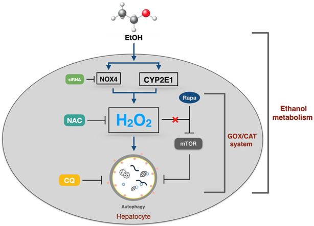
Alcoholic liver disease (ALD) is the most common liver disease worldwide and its underlying molecular mechanisms are still poorly understood. Moreover, conflicting data have been reported on potentially protective autophagy, the exact role of ethanol-metabolizing enzymes and ROS.
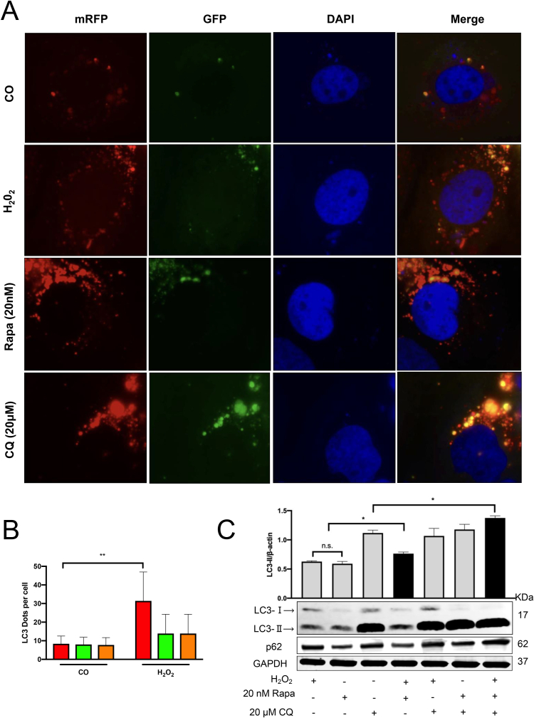
Expression of LC3B, CYP2E1, and NOX4 was studied in a mouse model of acute ethanol exposure by immunoblotting and immunohistochemistry. Autophagy was further studied in primary mouse hepatocytes and huh7 cells in response to ethanol and its major intermediator acetaldehyde. Experiments were carried out in cells overexpressing CYP2E1 and knock down of NOX4 using siRNA. The response to external HO was studied by using the GOX/CAT system. Autophagic flux was monitored using the mRFP-GFP-LC3 plasmid, while rapamycin and chloroquine served as positive and negative controls.
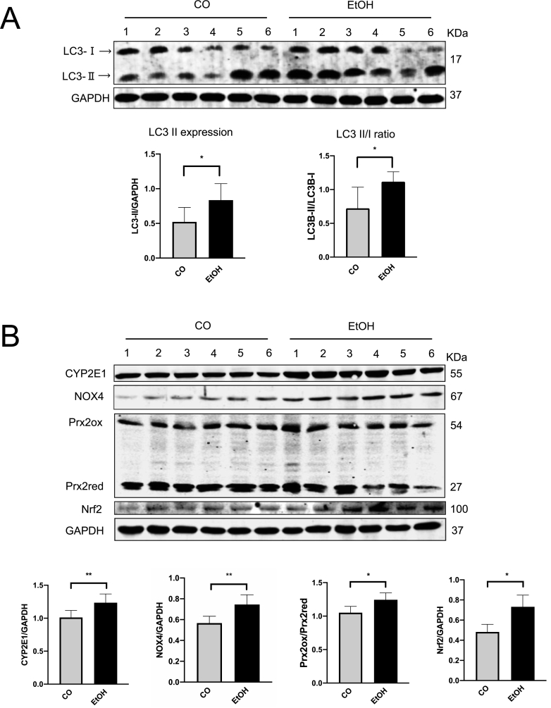
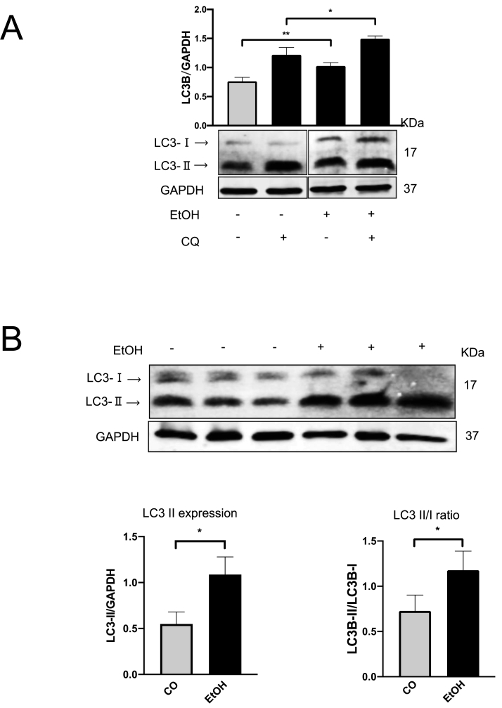
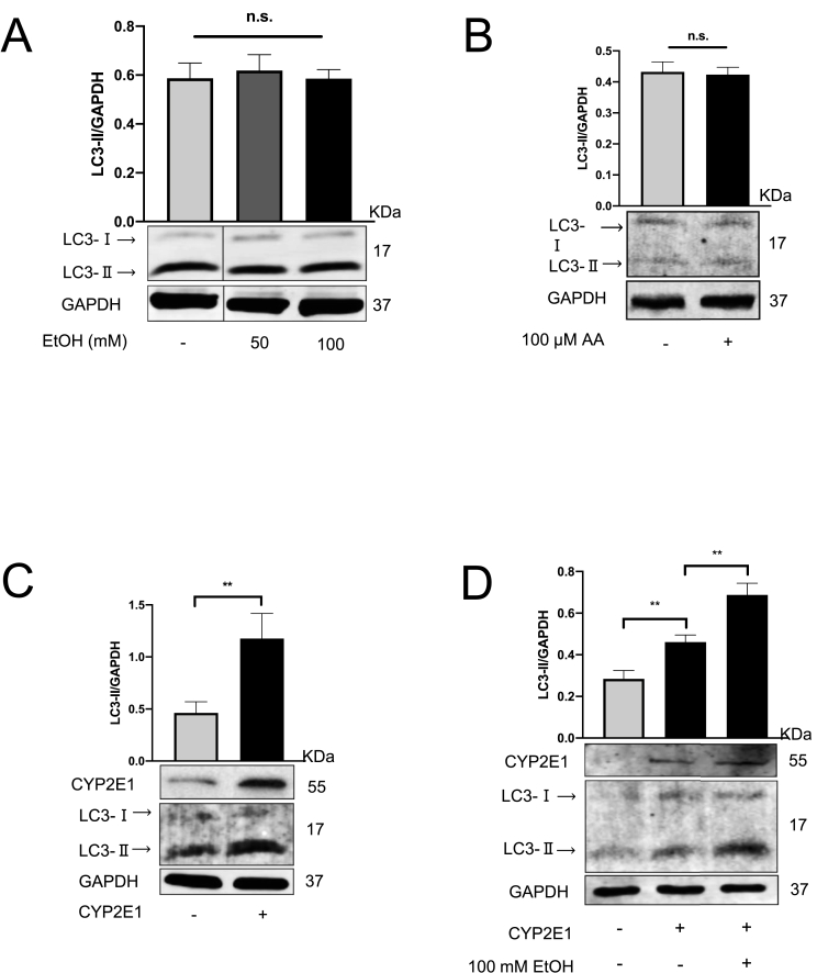
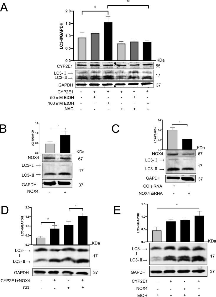
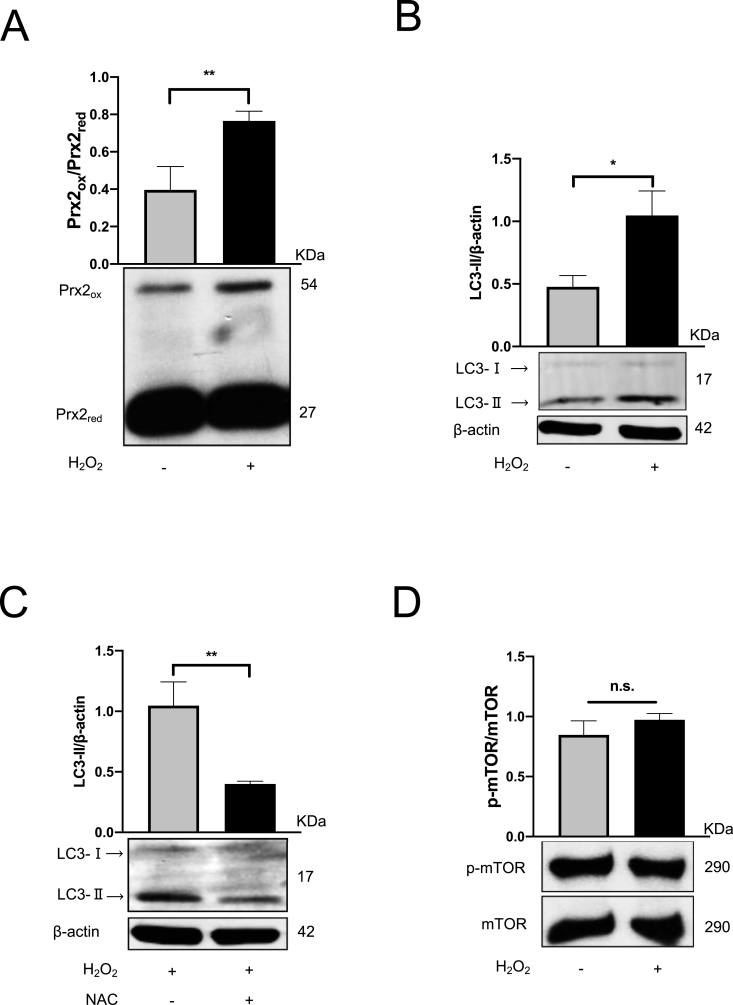
Acute ethanol exposure of mice over 24 h significantly induced autophagy as measured by LC3B expression but also induced the ROS-generating CYP2E1 and NOX4 enzymes. Notably, ethanol but not its downstream metabolite acetaldehyde induced autophagy in primary mouse hepatocytes. In contrast, autophagy could only be induced in huh7 cells in the presence of overexpressed CYP2E1. In addition, overexpression of NOX4 also significantly increased autophagy, which could be blocked by siRNA mediated knock down. The antioxidant N-acetylcysteine (NAC) also efficiently blocked CYP2E1-and NOX4-mediated induction of autophagy. Finally, specific and non-toxic production of HO by the GOX/CAT system as evidenced by elevated peroxiredoxin (Prx-2) also induced LC3B which was efficiently blocked by NAC. HO strongly increased the autophagic flux as measured by mRFP-GFP-LC3 plasmid.
We here provide evidence that short-term ethanol exposure induces autophagy in hepatocytes both in vivo and in vitro through the generation of ROS. These data suggest that suppression of autophagy by ethanol is most likely due to longer alcohol exposure during chronic alcohol consumption with the accumulation of e.g. misfolded proteins.
酒精性肝病(ALD)是全球最常见的肝脏疾病,但其潜在的分子机制仍知之甚少。此外,关于潜在的保护性自噬、乙醇代谢酶和 ROS 的确切作用,已有相互矛盾的数据报道。
通过免疫印迹和免疫组织化学研究了急性乙醇暴露小鼠模型中 LC3B、CYP2E1 和 NOX4 的表达。进一步研究了乙醇及其主要中间产物乙醛对原代小鼠肝细胞和 huh7 细胞的自噬作用。在过表达 CYP2E1 和用 siRNA 敲低 NOX4 的细胞中进行了实验。通过使用 GOX/CAT 系统研究了对外源性 HO 的反应。使用 mRFP-GFP-LC3 质粒监测自噬流,而雷帕霉素和氯喹分别作为阳性和阴性对照。
在 24 小时内,急性乙醇暴露显著诱导了 LC3B 表达所测量的自噬,但也诱导了产生 ROS 的 CYP2E1 和 NOX4 酶。值得注意的是,乙醇而不是其下游代谢产物乙醛诱导了原代小鼠肝细胞的自噬。相反,只有在过表达 CYP2E1 的情况下,huh7 细胞才能诱导自噬。此外,NOX4 的过表达也显著增加了自噬,而用 siRNA 介导的敲低可以阻断这种作用。抗氧化剂 N-乙酰半胱氨酸(NAC)也能有效地阻断 CYP2E1 和 NOX4 介导的自噬诱导。最后,GOX/CAT 系统产生的特异性、非毒性 HO,如过氧化物酶(Prx-2)升高所证明的,也诱导了 LC3B,而 NAC 则有效地阻断了 LC3B。HO 强烈增加了 mRFP-GFP-LC3 质粒测量的自噬流。
我们在此提供的证据表明,在体内和体外,短期乙醇暴露通过产生 ROS 诱导肝细胞自噬。这些数据表明,乙醇对自噬的抑制作用很可能是由于在慢性酒精摄入期间,酒精暴露时间较长,例如错误折叠的蛋白质积累所致。