National Cancer Center/National Clinical Research Center for Cancer/Cancer Hospital, Chinese Academy of Medical Sciences and Peking Union Medical College, Beijing, China.
Sleep Medicine Center, West China Hospital, Sichuan University, Chengdu, China.
Front Immunol. 2021 Jul 20;12:677169. doi: 10.3389/fimmu.2021.677169. eCollection 2021.
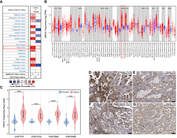
The endoplasmic reticulum oxidoreductin-1-like () gene encodes an endoplasmic reticulum luminal localized glycoprotein known to associated with hypoxia, however, the role of in shaping the tumor immune microenvironment (TIME) is yet to be elucidated in lung adenocarcinoma (LUAD).
In this study, raw datasets (including RNA-seq, methylation, sgRNA-seq, phenotype, and survival data) were obtained from public databases. This data was analyzed and used to explore the biological landscape of in immune infiltration. Expression data was used to characterize samples. Using gene signatures and cell quantification, stromal and immune infiltration was determined. These findings were used to predict sensitivity to immunotherapy.
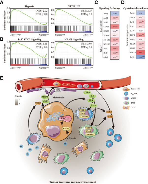
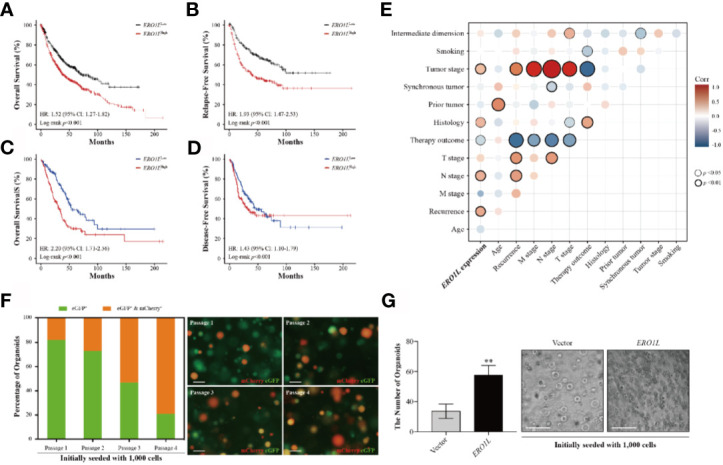
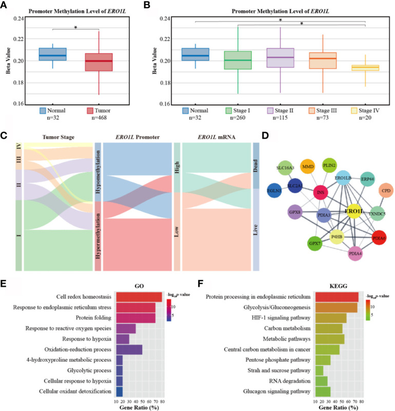
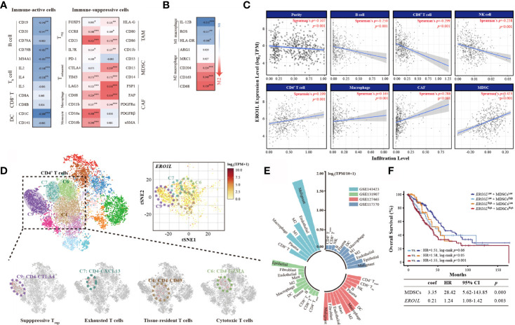
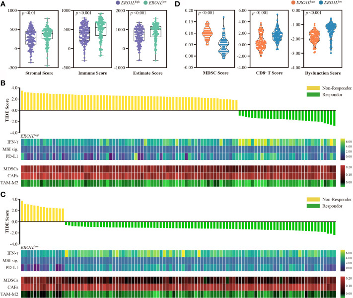
This study found that was significantly overexpressed in LUAD in comparison to normal tissue. This overexpression was found to be a result of hypomethylation of the promoter. Overexpression of resulted in an immune-suppressive TIME the recruitment of immune-suppressive cells including regulatory T cells (T), cancer associated fibroblasts, M2-type macrophages, and myeloid-derived suppressor cells. Using the Tumor Immune Dysfunction and Exclusion (TIDE) framework, it was identified that patients in the group possessed a significantly lower response rate to immunotherapy in comparison to the group. Mechanistic analysis revealed that overexpression of was associated with the upregulation of JAK-STAT and NF-κB signaling pathways, thus affecting chemokine and cytokine patterns in the TIME.
This study found that overexpression of was associated with poor prognoses in patients with LUAD. Overexpression of was indicative of a hypoxia-induced immune-suppressive TIME, which was shown to confer resistance to immunotherapy in patients with LUAD. Further studies are required to assess the potential role of as a biomarker for immunotherapy efficacy in LUAD.
内质网氧化还原酶 1 样()基因编码一种内质网腔定位的糖蛋白,已知与低氧有关,然而,在肺腺癌(LUAD)中,其在塑造肿瘤免疫微环境(TIME)中的作用尚未阐明。
本研究从公共数据库中获取了原始数据集(包括 RNA-seq、甲基化、sgRNA-seq、表型和生存数据)。对这些数据进行了分析,用于探索在免疫浸润中编码的生物学特征。表达数据用于对样本进行特征描述。利用基因特征和细胞定量,确定了基质和免疫浸润。这些发现被用于预测对免疫治疗的敏感性。
本研究发现与正常组织相比,在 LUAD 中显著过表达。这种过表达是由于启动子的低甲基化所致。过表达导致免疫抑制性 TIME 的招募,包括调节性 T 细胞(T)、癌症相关成纤维细胞、M2 型巨噬细胞和髓系来源的抑制细胞。利用肿瘤免疫功能障碍和排除(TIDE)框架,鉴定出在 组的患者对免疫治疗的反应率明显低于 组。机制分析表明,过表达与 JAK-STAT 和 NF-κB 信号通路的上调有关,从而影响 TIME 中的趋化因子和细胞因子模式。
本研究发现,在 LUAD 患者中,的过表达与不良预后相关。过表达与缺氧诱导的免疫抑制性 TIME 相关,表明其可导致 LUAD 患者对免疫治疗的耐药性。需要进一步的研究来评估作为 LUAD 免疫治疗疗效的潜在生物标志物的作用。