Jenkins Joshua, Ishak Mohd I, Eales Marcus, Gholinia Ali, Kulkarni Satishkumar, Keller Thomas F, May Paul W, Nobbs Angela H, Su Bo
Bristol Dental School, University of Bristol, Bristol, UK.
Faculty of Engineering Technology, Universiti Malaysia Perlis, Malaysia.
iScience. 2021 Jul 7;24(7):102818. doi: 10.1016/j.isci.2021.102818. eCollection 2021 Jul 23.
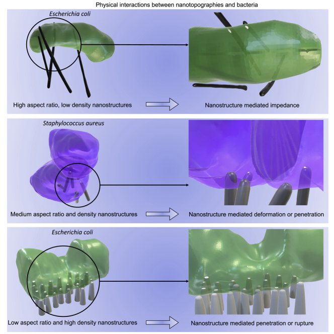
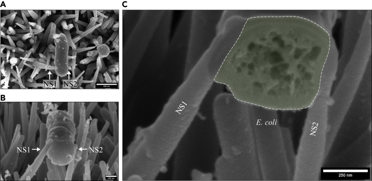
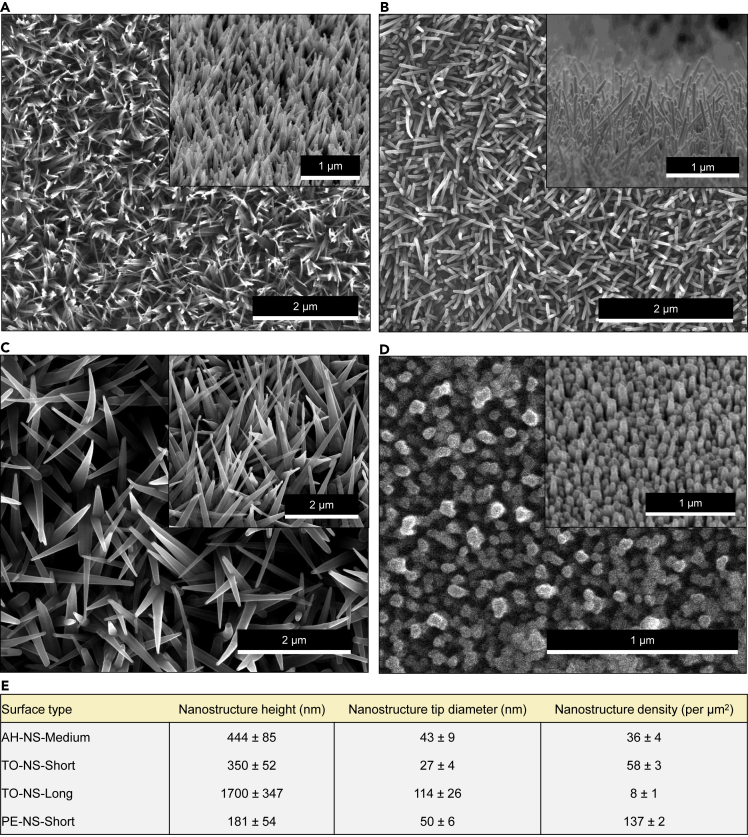
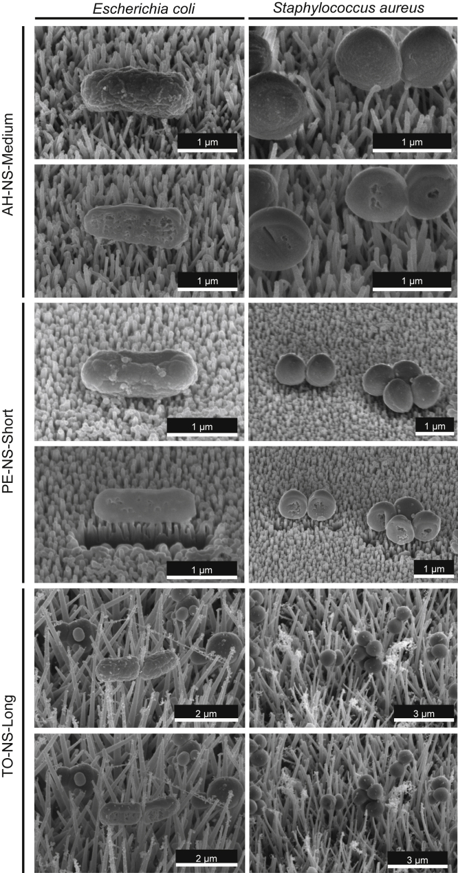
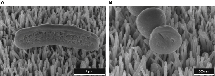
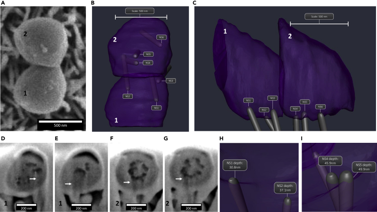
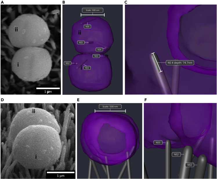
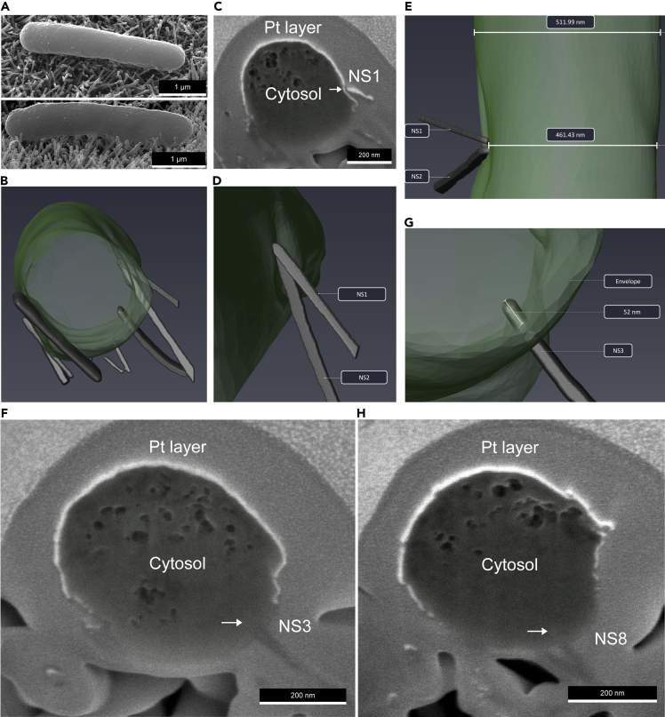
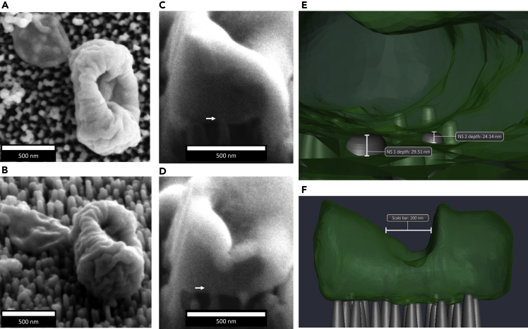
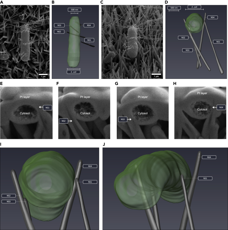
To robustly assess the antibacterial mechanisms of nanotopographies, it is critical to analyze the bacteria-nanotopography adhesion interface. Here, we utilize focused ion beam milling combined with scanning electron microscopy to generate three-dimensional reconstructions of or interacting with nanotopographies. For the first time, 3D morphometric analysis has been exploited to quantify the intrinsic contact area between each nanostructure and the bacterial envelope, providing an objective framework from which to derive the possible antibacterial mechanisms of synthetic nanotopographies. Surfaces with nanostructure densities between 36 and 58 per μm and tip diameters between 27 and 50 nm mediated envelope deformation and penetration, while surfaces with higher nanostructure densities (137 per μm) induced envelope penetration and mechanical rupture, leading to marked reductions in cell volume due to cytosolic leakage. On nanotopographies with densities of 8 per μm and tip diameters greater than 100 nm, bacteria predominantly adhered between nanostructures, resulting in cell impedance.
为了有力地评估纳米拓扑结构的抗菌机制,分析细菌与纳米拓扑结构的粘附界面至关重要。在此,我们利用聚焦离子束铣削结合扫描电子显微镜来生成与纳米拓扑结构相互作用的细菌的三维重建图像。首次利用三维形态计量分析来量化每个纳米结构与细菌包膜之间的固有接触面积,从而提供一个客观的框架,从中推导合成纳米拓扑结构可能的抗菌机制。纳米结构密度在每微米36至58个且尖端直径在27至50纳米之间的表面介导包膜变形和穿透,而纳米结构密度较高(每微米137个)的表面诱导包膜穿透和机械破裂,由于胞质泄漏导致细胞体积显著减小。在纳米结构密度为每微米8个且尖端直径大于100纳米的纳米拓扑结构上,细菌主要粘附在纳米结构之间,导致细胞阻抗。