Kukreja Bhavna Jha, Bhat Kishore Gajanan, Kukreja Pankaj, Kumber Vijay Mahadev, Balakrishnan Rajkumar, Govila Vivek
Department of Periodontology, Babu Banarasi Das College of Dental Sciences, Babu Banarasi Das University, Lucknow, Uttar Pradesh, India.
Department of Microbiology, Maratha Mandal's Nathajirao G. Halgekar Institute of Dental Sciences and Research Centre, Belagavi, Karnataka, India.
J Indian Soc Periodontol. 2021 Jul-Aug;25(4):295-299. doi: 10.4103/jisp.jisp_442_20. Epub 2021 Jul 1.
It is a known fact that periodontal tissue regeneration can be achieved by the use of periodontal ligament stem cells (PDLSCs). Current mainstay of periodontal treatment is focusing on stem cell tissue engineering as an effective therapy, making it important to isolate PDLSCs from periodontal tissues.
The present research endeavor was undertaken to elucidate a technique for isolating PDLSCs for reconstructing the natural PDL tissue.
The study design involves prospective study.
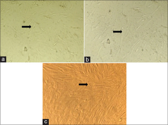
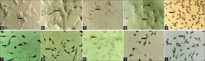
Premolar teeth were extracted from 12 patients who were under orthodontic treatment. PDL cells were scraped from their roots. Using 10 ml of Dulbecco's modified Eagle's medium with pH 7.2, the specimens of the periodontal tissue were transferred to laboratory where cell culture was done. Isolated stem cells were grown on 24-well microtiter plates-containing cover slips. They were incubated overnight at approximately 37°C in 95% air and 5% humidification. Anti-CD 45, CD73, CD90, CD105, and CD146 antibodies were used. After staining, cells were observed under phase-contrast microscopy and in inverted microscope.
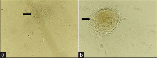
The cells showed a marked growth and 90% confluence at day 6. Cells presented thin and long fibroblastic spindle morphology. Isolated PDLSCs showed colony-forming ability at the 14 day after seeding. Immunohistochemical staining of PDLSCs showed positive uptake for CD146, CD90, CD73, CD105, and negative uptake for CD45.
The human PDLSCs can be clearly isolated and characterized by using CD90, CD73, CD146, and CD105 markers of stem cells.
众所周知,使用牙周膜干细胞(PDLSCs)可实现牙周组织再生。牙周治疗的当前主要方法是将干细胞组织工程作为一种有效疗法,因此从牙周组织中分离PDLSCs很重要。
本研究旨在阐明一种分离PDLSCs以重建天然牙周膜组织的技术。
该研究设计为前瞻性研究。
从12名接受正畸治疗的患者中拔除前磨牙。从牙根刮取牙周膜细胞。使用10毫升pH值为7.2的杜氏改良 Eagle 培养基,将牙周组织标本转移至实验室进行细胞培养。分离出的干细胞在含有盖玻片的24孔微量滴定板上生长。它们在约37°C、95%空气和5%湿度的条件下孵育过夜。使用抗CD45、CD73、CD90、CD105和CD146抗体。染色后,在相差显微镜和倒置显微镜下观察细胞。
细胞在第6天显示出显著生长且汇合度达90%。细胞呈现细长的成纤维细胞纺锤形形态。接种后第14天,分离出的PDLSCs显示出集落形成能力。PDLSCs的免疫组织化学染色显示CD146、CD90、CD73、CD105呈阳性摄取,CD45呈阴性摄取。
使用干细胞的CD90、CD73、CD146和CD105标记物可清晰分离和鉴定人PDLSCs。