Kats Lazar, Goldman Yuli, Kahn Adrian
Department of Oral Pathology, Oral Medicine and Maxillofacial Imaging, School of Dental Medicine, Tel Aviv University, 69978, Tel Aviv, Israel.
Department of Oral and Maxillofacial Surgery, School of Dental Medicine, Tel Aviv University, Tel Aviv, Israel.
BMC Oral Health. 2021 Aug 19;21(1):411. doi: 10.1186/s12903-021-01777-9.
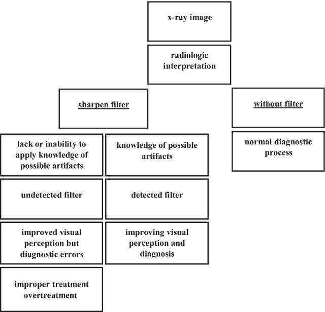
Improvement of image quality in radiology, including the maxillofacial region, is important for diagnosis by enhancing the visual perception of the original image. One of the most used modification methods is sharpening, in which simultaneously with the improvement, due to edge enhancement, several artifacts appear. These might lead to misdiagnosis and, as a consequence, to improper treatment. The purpose of this study was to prove the feasibility and effectiveness of automatic sharpening detection based on neural networks.
The in-house created dataset contained 4290 X-ray slices from different datasets of cone beam computed tomography images were taken on 2 different devices: Ortophos 3D SL (Sirona Dental Systems GmbH, Bensheim, Germany) and Planmeca ProMax 3D (Planmeca, Helsinki, Finland). The selected slices were modified using the sharpening filter available in the software RadiAnt Dicom Viewer software (Medixant, Poland), version 5.5. The neural network known as "ResNet-50" was used, which has been previously trained on the ImageNet dataset. The input images and their corresponding sharpening maps were used to train the network. For the implementation, Keras with Tensorflow backend was used. The model was trained using NVIDIA GeForce GTX 1080 Ti GPU. Receiver Operating Characteristic (ROC) analysis was performed to calculate the detection accuracy using MedCalc Statistical Software version 14.8.1 (MedCalc Software Ltd, Ostend, Belgium). The study was approved by the Ethical Committee.
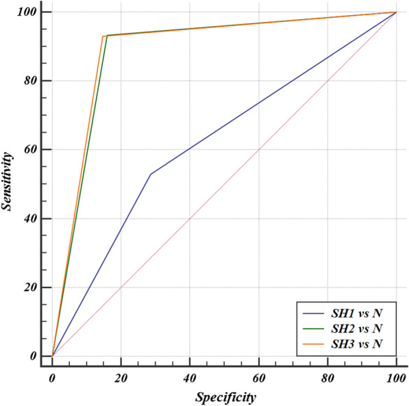
For the test, 1200 different images with the filter and without modification were used. An analysis of the detection of three different levels of sharpening (1, 2, 3) showed sensitivity of 53%, 93.33%, 93% and specificity of 72.33%, 84%, 85.33%, respectively with an accuracy of 62.17%, 88.67% and 89% (p < 0.0001). The ROC analysis in all tests showed an Area Under Curve (AUC) different from 0.5 (null hypothesis).
This study showed a high performance in automatic sharpening detection of radiological images based on neural network technology. Further investigation of these capabilities, including their application to different types of radiological images, will significantly improve the level of diagnosis and appropriate treatment.
提高包括颌面区域在内的放射学图像质量,对于通过增强原始图像的视觉感知来进行诊断非常重要。最常用的修改方法之一是锐化,在提高图像质量的同时,由于边缘增强会出现一些伪影。这些伪影可能导致误诊,进而导致治疗不当。本研究的目的是证明基于神经网络的自动锐化检测的可行性和有效性。
内部创建的数据集包含来自不同锥束计算机断层扫描图像数据集的4290张X线切片,这些图像是在2种不同设备上采集的:Ortophos 3D SL(德国本斯海姆的西诺德牙科系统有限公司)和Planmeca ProMax 3D(芬兰赫尔辛基的普兰梅卡公司)。使用RadiAnt Dicom Viewer软件(波兰的Medixant公司)5.5版本中可用的锐化滤镜对所选切片进行修改。使用名为“ResNet-50”的神经网络,该网络先前已在ImageNet数据集上进行过训练。输入图像及其相应的锐化图用于训练网络。在实现过程中,使用了以TensorFlow为后端的Keras。该模型使用NVIDIA GeForce GTX 1080 Ti GPU进行训练。使用MedCalc统计软件14.8.1版本(比利时奥斯坦德的MedCalc软件有限公司)进行受试者工作特征(ROC)分析,以计算检测准确率。该研究获得了伦理委员会的批准。
在测试中,使用了1200张经过滤镜处理和未修改的不同图像。对三种不同锐化水平(1、2、3)的检测分析显示,敏感性分别为53%、93.33%、93%,特异性分别为72.33%、84%、85.33%,准确率分别为62.17%、88.67%和89%(p < 0.0001)。所有测试中的ROC分析显示曲线下面积(AUC)均不同于0.5(零假设)。
本研究表明基于神经网络技术的放射学图像自动锐化检测具有高性能。对这些能力的进一步研究,包括将其应用于不同类型的放射学图像,将显著提高诊断水平和恰当治疗水平。