• 文献检索
  • 文档翻译
  • 深度研究
  • 学术资讯
  • Suppr Zotero 插件Zotero 插件
  • 邀请有礼
  • 套餐&价格
  • 历史记录
应用&插件
Suppr Zotero 插件Zotero 插件浏览器插件Mac 客户端Windows 客户端微信小程序
定价
高级版会员购买积分包购买API积分包
服务
文献检索文档翻译深度研究API 文档MCP 服务
关于我们
关于 Suppr公司介绍联系我们用户协议隐私条款
关注我们

Suppr 超能文献

核心技术专利:CN118964589B侵权必究
粤ICP备2023148730 号-1Suppr @ 2026

文献检索

告别复杂PubMed语法,用中文像聊天一样搜索,搜遍4000万医学文献。AI智能推荐,让科研检索更轻松。

立即免费搜索

文件翻译

保留排版,准确专业,支持PDF/Word/PPT等文件格式,支持 12+语言互译。

免费翻译文档

深度研究

AI帮你快速写综述,25分钟生成高质量综述,智能提取关键信息,辅助科研写作。

立即免费体验

BO-CLAHE增强新生儿胸部X光图像质量以改善病变分类。

BO-CLAHE enhancing neonatal chest X-ray image quality for improved lesion classification.

作者信息

Han Jiwon, Choi Byungmin, Kim Jae Young, Lee Yeonjoon

机构信息

Department of Applied Artificial Intelligence, Hanyang University, Seoul, 15588, Korea.

Department of Pediatrics, Ansan Hospital, College of Medicine, Korea University, Ansan, 15355, Republic of Korea.

出版信息

Sci Rep. 2025 Feb 10;15(1):4931. doi: 10.1038/s41598-025-88451-0.

DOI:10.1038/s41598-025-88451-0
PMID:39929905
原文链接:https://pmc.ncbi.nlm.nih.gov/articles/PMC11811177/
Abstract

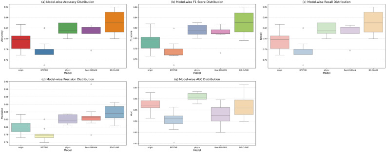
In the case of neonates, especially low birth weight preterm and high-risk infants, portable X-rays are frequently used. However, the image quality of portable X-rays is significantly lower compared to standard adult or pediatric X-rays, leading to considerable challenges in identifying abnormalities. Although attempts have been made to introduce deep learning to address these image quality issues, the poor quality of the images themselves hinders the training of deep learning models, further emphasizing the need for image enhancement. Additionally, since neonates have a high cell division rate and are highly sensitive to radiation, increasing radiation exposure to improve image quality is not a viable solution. Therefore, it is crucial to enhance image quality through preprocessing before training deep learning models. While various image enhancement methods have been proposed, Contrast Limited Adaptive Histogram Equalization (CLAHE) has been recognized as an effective technique for contrast-based image improvement. However, despite extensive research, the process of setting CLAHE's hyperparameters still relies on a brute force, manual approach, making it inefficient. To address this issue, we propose a method called Bayesian Optimization CLAHE(BO-CLAHE), which leverages Bayesian optimization to automatically select the optimal hyperparameters for X-ray images used in diagnosing lung diseases in preterm and high-risk neonates. The images enhanced by BO-CLAHE demonstrated superior performance across several classification models, with particularly notable improvements in diagnosing Transient Tachypnea of the Newborn (TTN). This approach not only reduces radiation exposure but also contributes to the development of AI-based diagnostic tools, playing a crucial role in the early diagnosis and treatment of preterm and high-risk neonates.

摘要

对于新生儿,尤其是低出生体重早产儿和高危婴儿,便携式X射线经常被使用。然而,与标准的成人或儿科X射线相比,便携式X射线的图像质量明显较低,这给识别异常带来了相当大的挑战。尽管已经尝试引入深度学习来解决这些图像质量问题,但图像本身的质量较差阻碍了深度学习模型的训练,进一步凸显了图像增强的必要性。此外,由于新生儿的细胞分裂率高且对辐射高度敏感,增加辐射暴露以提高图像质量不是一个可行的解决方案。因此,在训练深度学习模型之前通过预处理提高图像质量至关重要。虽然已经提出了各种图像增强方法,但对比度受限自适应直方图均衡化(CLAHE)已被公认为是一种基于对比度改善图像的有效技术。然而,尽管进行了广泛的研究,设置CLAHE超参数的过程仍然依赖于强力的手动方法,效率低下。为了解决这个问题,我们提出了一种名为贝叶斯优化CLAHE(BO-CLAHE)的方法,该方法利用贝叶斯优化自动为用于诊断早产儿和高危新生儿肺部疾病的X射线图像选择最佳超参数。通过BO-CLAHE增强的图像在多个分类模型中表现出卓越的性能,在诊断新生儿暂时性呼吸急促(TTN)方面有特别显著的改进。这种方法不仅减少了辐射暴露,还为基于人工智能的诊断工具的开发做出了贡献,在早产儿和高危新生儿的早期诊断和治疗中发挥着关键作用。

https://cdn.ncbi.nlm.nih.gov/pmc/blobs/311f/11811177/22c167a5935b/41598_2025_88451_Fig8_HTML.jpg
https://cdn.ncbi.nlm.nih.gov/pmc/blobs/311f/11811177/a7d7460ebadc/41598_2025_88451_Fig1_HTML.jpg
https://cdn.ncbi.nlm.nih.gov/pmc/blobs/311f/11811177/3597bbc9cdfe/41598_2025_88451_Fig2_HTML.jpg
https://cdn.ncbi.nlm.nih.gov/pmc/blobs/311f/11811177/f694ff19d079/41598_2025_88451_Fig3_HTML.jpg
https://cdn.ncbi.nlm.nih.gov/pmc/blobs/311f/11811177/68fdfc9f96c9/41598_2025_88451_Fig4_HTML.jpg
https://cdn.ncbi.nlm.nih.gov/pmc/blobs/311f/11811177/8267123ef63d/41598_2025_88451_Fig5_HTML.jpg
https://cdn.ncbi.nlm.nih.gov/pmc/blobs/311f/11811177/d105b03aab41/41598_2025_88451_Fig6_HTML.jpg
https://cdn.ncbi.nlm.nih.gov/pmc/blobs/311f/11811177/6935ee2287db/41598_2025_88451_Figa_HTML.jpg
https://cdn.ncbi.nlm.nih.gov/pmc/blobs/311f/11811177/c028ef0aef05/41598_2025_88451_Fig7_HTML.jpg
https://cdn.ncbi.nlm.nih.gov/pmc/blobs/311f/11811177/22c167a5935b/41598_2025_88451_Fig8_HTML.jpg

相似文献

1
BO-CLAHE enhancing neonatal chest X-ray image quality for improved lesion classification.BO-CLAHE增强新生儿胸部X光图像质量以改善病变分类。
Sci Rep. 2025 Feb 10;15(1):4931. doi: 10.1038/s41598-025-88451-0.
2
Enhancing semantic segmentation in chest X-ray images through image preprocessing: ps-KDE for pixel-wise substitution by kernel density estimation.通过图像预处理增强胸部 X 光图像的语义分割:基于核密度估计的像素级替换的 ps-KDE。
PLoS One. 2024 Jun 24;19(6):e0299623. doi: 10.1371/journal.pone.0299623. eCollection 2024.
3
Automatic x-ray image contrast enhancement based on parameter auto-optimization.基于参数自动优化的X射线图像自动对比度增强
J Appl Clin Med Phys. 2017 Nov;18(6):218-223. doi: 10.1002/acm2.12172. Epub 2017 Sep 6.
4
Pulmonary abnormality screening on chest x-rays from different machine specifications: a generalized AI-based image manipulation pipeline.不同机型胸片肺部异常筛查:基于 AI 的通用图像操作流程。
Eur Radiol Exp. 2023 Nov 9;7(1):68. doi: 10.1186/s41747-023-00386-1.
5
COVID-19 detection from chest X-ray images using CLAHE-YCrCb, LBP, and machine learning algorithms.使用 CLAHE-YCrCb、LBP 和机器学习算法从胸部 X 光图像中检测 COVID-19。
BMC Bioinformatics. 2024 Jan 17;25(1):28. doi: 10.1186/s12859-023-05427-5.
6
Enhancing Medical Image Classification through Transfer Learning and CLAHE Optimization.通过迁移学习和对比度受限自适应直方图均衡化(CLAHE)优化增强医学图像分类
Curr Med Imaging. 2025;21:e15734056342623. doi: 10.2174/0115734056342623241119061744.
7
Evaluation of histogram equalization and contrast limited adaptive histogram equalization effect on image quality and fractal dimensions of digital periapical radiographs.评价直方图均衡化和限制对比度自适应直方图均衡化对数字根尖射线照片的图像质量和分形维数的影响。
Oral Radiol. 2023 Apr;39(2):418-424. doi: 10.1007/s11282-022-00654-7. Epub 2022 Sep 8.
8
SVD-CLAHE boosting and balanced loss function for Covid-19 detection from an imbalanced Chest X-Ray dataset.SVD-CLAHE 增强和平衡损失函数在不平衡的 Chest X-Ray 数据集上进行 Covid-19 检测。
Comput Biol Med. 2022 Nov;150:106092. doi: 10.1016/j.compbiomed.2022.106092. Epub 2022 Sep 28.
9
Enhanced Pneumonia Detection in Chest X-Rays Using Hybrid Convolutional and Vision Transformer Networks.使用混合卷积和视觉Transformer网络增强胸部X光片中的肺炎检测
Curr Med Imaging. 2025;21:e15734056326685. doi: 10.2174/0115734056326685250101113959.
10
Explainable hybrid transformer for multi-classification of lung disease using chest X-rays.用于使用胸部X光进行肺部疾病多分类的可解释混合变压器。
Sci Rep. 2025 Feb 24;15(1):6650. doi: 10.1038/s41598-025-90607-x.

引用本文的文献

1
Diagnosis of pneumonia from chest X-ray images using YOLO deep learning.使用YOLO深度学习技术从胸部X光图像中诊断肺炎
Front Neurorobot. 2025 Apr 28;19:1576438. doi: 10.3389/fnbot.2025.1576438. eCollection 2025.

本文引用的文献

1
Automatic detection of image sharpening in maxillofacial radiology.颌面放射学中图像锐化的自动检测
BMC Oral Health. 2021 Aug 19;21(1):411. doi: 10.1186/s12903-021-01777-9.
2
Optimising the use of computed radiography in pediatric chest imaging.优化计算机X线摄影在儿科胸部成像中的应用。
J Digit Imaging. 2009 Apr;22(2):104-13. doi: 10.1007/s10278-007-9071-2. Epub 2007 Sep 13.
3
Missed non-small cell lung cancer: radiographic findings of potentially resectable lesions evident only in retrospect.漏诊的非小细胞肺癌:仅在回顾时才明显可见的潜在可切除病变的影像学表现。
Radiology. 2003 Jan;226(1):235-41. doi: 10.1148/radiol.2261011924.