Animal Biotechnology Division, National Institute of Animal Science, Rural Development Administration, Wanju 55365, Korea.
J Vet Sci. 2021 Sep;22(5):e63. doi: 10.4142/jvs.2021.22.e63. Epub 2021 Jul 14.
Recently, mesenchymal stem cells therapy has been performed in dogs, although the outcome is not always favorable.
To investigate the therapeutic efficacy of mesenchymal stem cells (MSCs) using dog leukocyte antigen (DLA) matching between the donor and recipient .
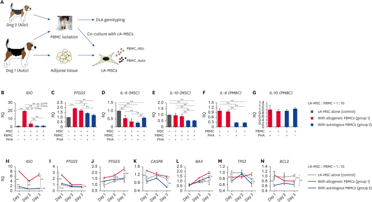
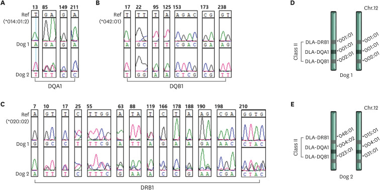
Canine adipose-derived MSCs (cA-MSCs) isolated from the subcutaneous tissue of Dog 1 underwent characterization. For major DLA genotyping (DQA1, DQB1, and DRB1), peripheral blood mononuclear cells (PBMCs) from two dogs (Dogs 1 and 2) were analyzed by direct sequencing of polymerase chain reaction (PCR) products. The cA-MSCs were co-cultured at a 1:10 ratio with activated PBMCs (DLA matching or mismatching) for 3 days and analyzed for immunosuppressive (, , and ), inflammatory ( and ), and apoptotic genes (, , , and ) by quantitative real-time reverse transcriptase-PCR.
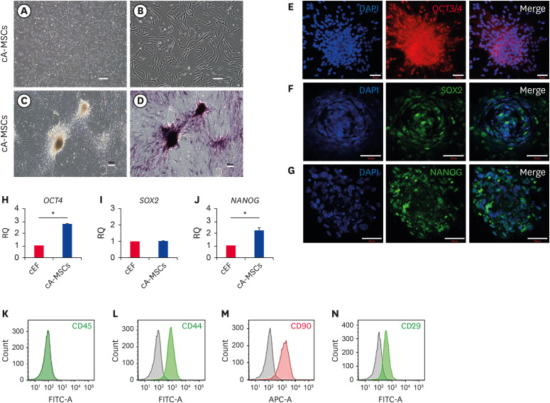
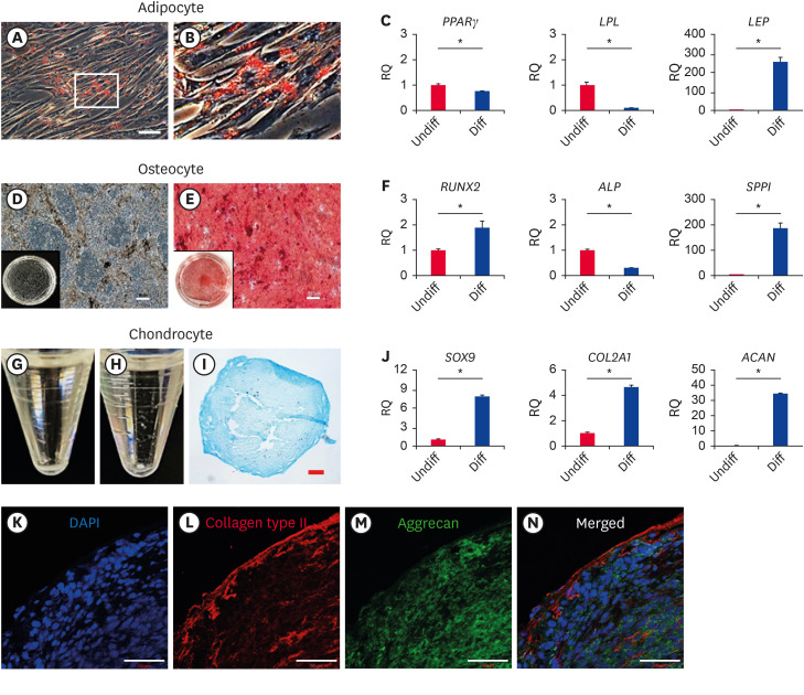
cA-MSCs were expressed cell surface markers such as CD90/44/29/45 and differentiated into osteocytes, chondrocytes, and adipocytes According to the Immuno Polymorphism Database, DLA genotyping comparisons of Dogs 1 and 2 revealed complete differences in genes DQA1, DQB1, and DRB1. In the co-culturing of cA-MSCs and PBMCs, DLA mismatch between the two cell types induced a significant increase in the expression of immunosuppressive () and apoptotic ) genes.
The administration of cA-MSCs matching the recipient DLA type can alleviate the need to regulate excessive immunosuppressive responses associated with genes, such as and Furthermore, easy and reliable DLA genotyping technology is required because of the high degree of genetic polymorphisms of DQA1, DQB1, and DRB1 and the low readability of DLA 88.
最近,间充质干细胞治疗已在犬中进行,尽管结果并不总是令人满意。
研究供体和受者之间犬白细胞抗原(DLA)匹配的间充质干细胞(MSCs)治疗的疗效。
从犬 1 的皮下组织分离出犬脂肪来源间充质干细胞(cA-MSCs),并对其进行特征描述。对两只犬(犬 1 和犬 2)的外周血单个核细胞(PBMCs)进行主要 DLA 基因分型(DQA1、DQB1 和 DRB1),通过聚合酶链反应(PCR)产物的直接测序进行分析。将 cA-MSCs 与激活的 PBMCs(DLA 匹配或不匹配)以 1:10 的比例共培养 3 天,并通过定量实时逆转录 PCR 分析免疫抑制(、、和)、炎症(和)和凋亡基因(、、、和)。
cA-MSCs 表达细胞表面标志物,如 CD90/44/29/45,并分化为成骨细胞、软骨细胞和脂肪细胞。根据免疫多态性数据库,犬 1 和犬 2 的 DLA 基因分型比较显示,基因 DQA1、DQB1 和 DRB1 完全不同。在 cA-MSCs 和 PBMCs 的共培养中,两种细胞类型之间的 DLA 不匹配诱导免疫抑制()和凋亡()基因的表达显著增加。
给予与受者 DLA 类型匹配的 cA-MSCs 可以减轻与基因相关的过度免疫抑制反应的需要,如和。此外,由于 DQA1、DQB1 和 DRB1 的遗传多态性程度高,DLA 88 的可读性低,因此需要易于且可靠的 DLA 基因分型技术。