Li Haicheng, Li Ting, Wang Heting, He Xuemin, Li Ying, Wen Siying, Peng Rongdong, Nie Yuanpeng, Lu Yan, Yang He, Ye Yinong, Shi Guojun, Chen Yanming
Department of Endocrinology and Metabolism, The Third Affiliated Hospital of Sun Yat-Sen University, Guangzhou, China.
Cancer Science Institute of Singapore, National University of Singapore, Singapore, Singapore.
Front Cardiovasc Med. 2021 Aug 11;8:689318. doi: 10.3389/fcvm.2021.689318. eCollection 2021.
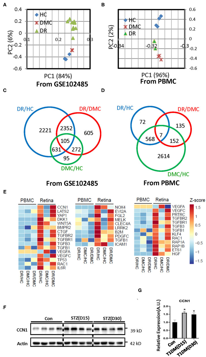
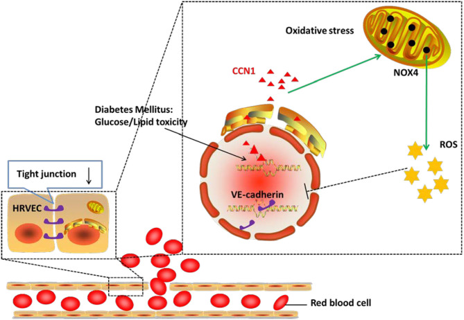
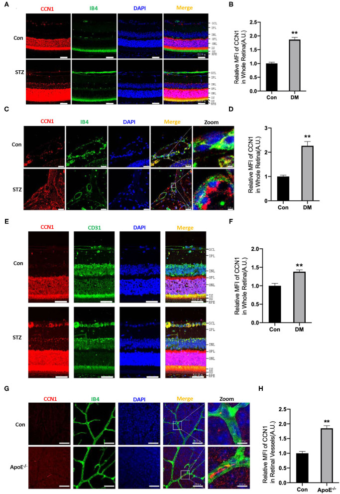
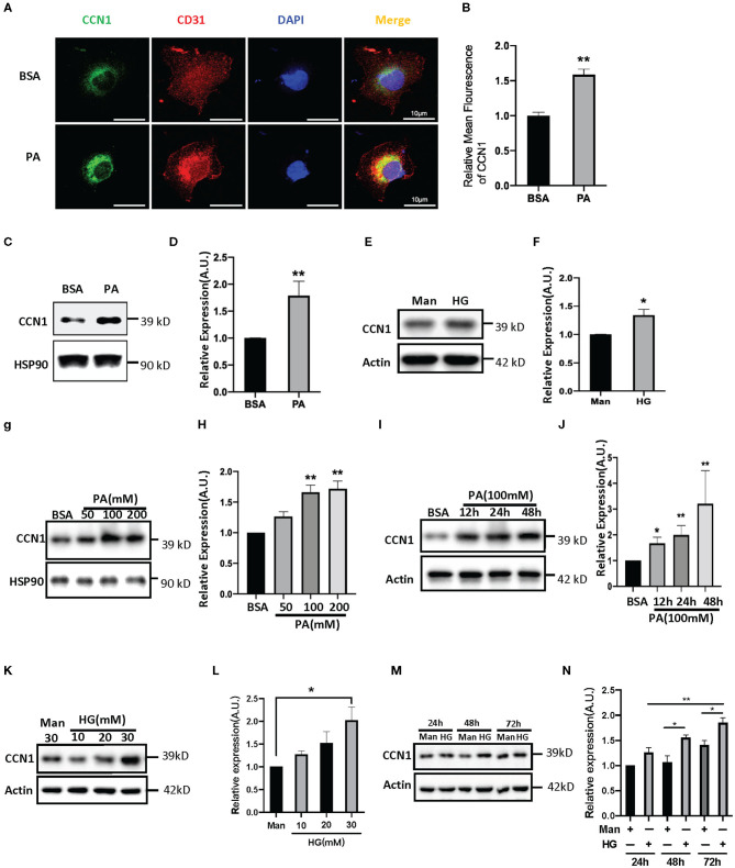
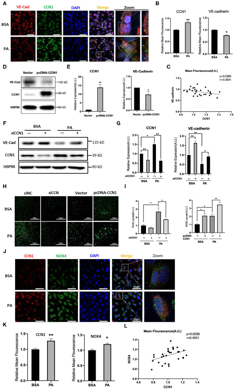
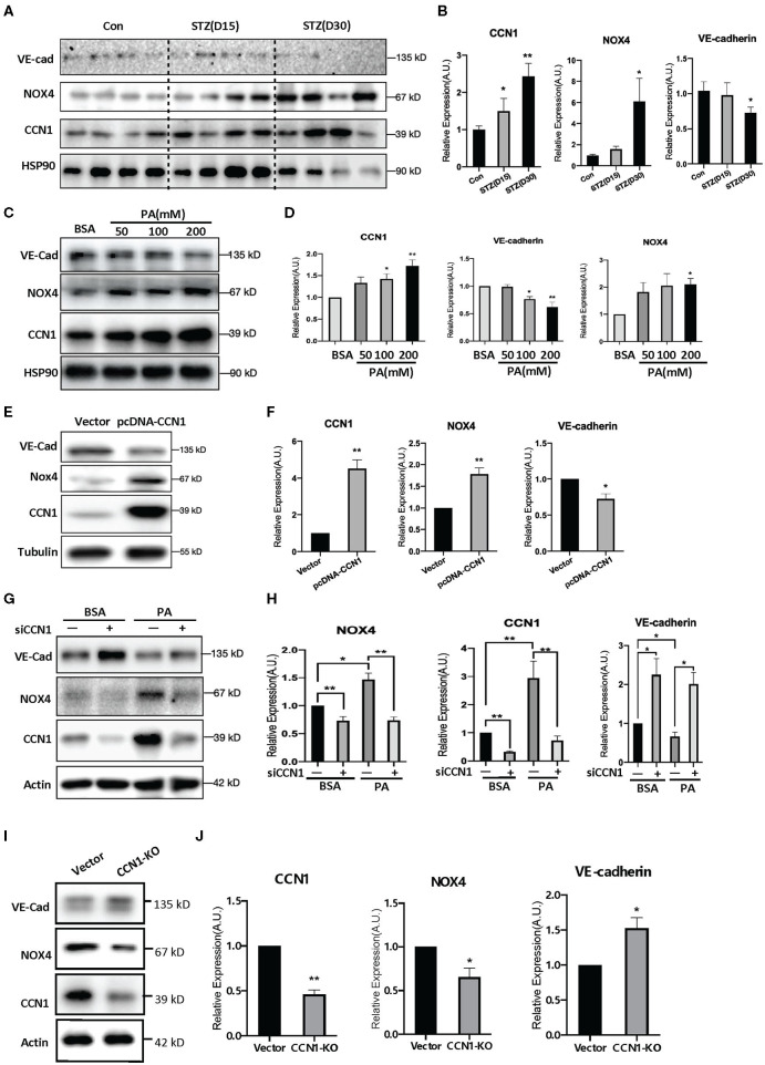
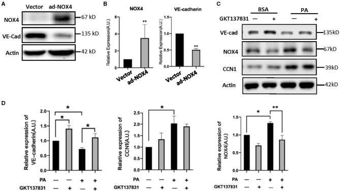
Diabetic retinopathy (DR) is one of the most common diabetic microvascular complications. However, the pathogenesis of DR has not yet been fully elucidated. This study aimed to discover novel and key molecules involved in the pathogenesis of DR, which could potentially be targets for therapeutic DR intervention. To identify potential genes involved in the pathogenesis of DR, we analyzed the public database of neovascular membranes (NVMs) from patients with proliferative diabetic retinopathy (PDR) and healthy controls (HCs) (GSE102485, https://www.ncbi.nlm.nih.gov/geo/query/acc.cgi?acc=GSE102485). Further, we compared these findings by performing RNA-sequencing analysis of peripheral blood mononuclear cells (PBMC) from patients with DR, control patients with non-complicated diabetes mellitus (DMC), and HCs. To determine the critical role of candidate genes in DR, knockdown or knockout was performed in human retinal vascular endothelial cells (HRVECs). The oxidative stress pathway, as well as tight junction integrity, was analyzed. Transcriptional profiles showed distinct patterns between the NVMs of patients with DR and those of the HCs. Those genes enriched in either extracellular matrix (ECM)-receptor interaction or focal adhesion pathways were considerably upregulated. Both pathways were important for maintaining the integrity of retinal vascular structure and function. Importantly, the gene encoding the matricellular protein CCN1, a key gene in cell physiology, was differentially expressed in both pathways. Knockdown of CCN1 by small interfering RNA (siRNA) or knockout of CCN1 by the CRISPR-Cas9 technique in HRVECs significantly increased the levels of VE-cadherin, reduced the level of NADPH oxidase 4 (NOX4), and inhibited the generation of reactive oxygen species (ROS). The present study identifies CCN1 as an important regulator in the pathogenesis of DR. Increased expression of CCN1 stimulates oxidative stress and disrupts tight junction integrity in endothelial cells by inducing NOX4. Thus, targeting the CCN1/NOX4 axis provides a therapeutic strategy for treating DR by alleviating endothelial cell injury.
糖尿病视网膜病变(DR)是最常见的糖尿病微血管并发症之一。然而,DR的发病机制尚未完全阐明。本研究旨在发现参与DR发病机制的新的关键分子,这些分子可能成为DR治疗干预的靶点。为了鉴定参与DR发病机制的潜在基因,我们分析了增殖性糖尿病视网膜病变(PDR)患者和健康对照(HC)的新生血管膜(NVM)公共数据库(GSE102485,https://www.ncbi.nlm.nih.gov/geo/query/acc.cgi?acc=GSE102485)。此外,我们通过对DR患者、非复杂性糖尿病(DMC)对照患者和HC的外周血单个核细胞(PBMC)进行RNA测序分析来比较这些发现。为了确定候选基因在DR中的关键作用,我们在人视网膜血管内皮细胞(HRVEC)中进行了基因敲低或敲除。分析了氧化应激途径以及紧密连接的完整性。转录谱显示DR患者的NVM与HC的NVM之间存在明显差异。那些在细胞外基质(ECM)-受体相互作用或粘着斑途径中富集的基因显著上调。这两条途径对于维持视网膜血管结构和功能的完整性都很重要。重要的是,编码基质细胞蛋白CCN1的基因是细胞生理学中的关键基因,在这两条途径中均有差异表达。在HRVEC中通过小干扰RNA(siRNA)敲低CCN1或通过CRISPR-Cas9技术敲除CCN1可显著增加VE-钙黏蛋白水平,降低NADPH氧化酶4(NOX4)水平,并抑制活性氧(ROS)的产生。本研究确定CCN1是DR发病机制中的重要调节因子。CCN1表达增加会刺激氧化应激,并通过诱导NOX4破坏内皮细胞的紧密连接完整性。因此,靶向CCN1/NOX4轴为通过减轻内皮细胞损伤来治疗DR提供了一种治疗策略。