Precision Medicine Center, Taizhou Central Hospital (Taizhou University Hospital), Taizhou, 318000, Zhejiang, People's Republic of China.
College of Lab Medicine, Hebei North University, No.11, Zuanshi Road (south), Gaoxin District, Zhangjiakou, 075000, Hebei, People's Republic of China.
Diagn Pathol. 2021 Aug 31;16(1):84. doi: 10.1186/s13000-021-01139-7.
The incidence of papillary thyroid carcinoma (PTC) has been steadily increasing over the past decades. Hashimoto's thyroiditis (HT) is the most common autoimmune disease, and is related to the pathogenesis of PTC. Programmed death-1 (PD-1) is currently used for the treatment of PTC, but there are very few studies on the clinical value of PD-1 in the diagnosis and targeted therapy of PTC.
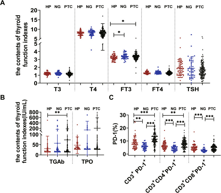
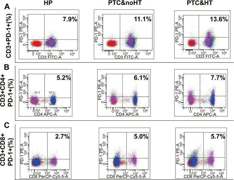
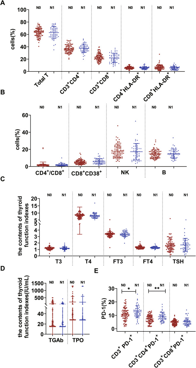
The expression of T, B, NK cells and PD-1 in the peripheral blood of 132 patients with PTC (PTC group), 48 patients with nodular goiter (NG group) and 63 healthy subjects (HP group) were detected by flow cytometry. The expression of plasma T3, T4, FT3, FT4, TSH, TGAb and TPO was detected by chemiluminescence immunoassay. Among 132 PTC, 49 PTC&HT and 83 PTC&noHT were included. Among 48 NG, 10 NG&HT and 38 NG&noHT were included. The expressions of programmed death- ligand1(PD-L1) in tumor tissues of PTC group and thyroid tissues of NG group, PD-1 and CD3 in tumor infiltration lymphocyte (TIL) were detected by immunohistochemistry.
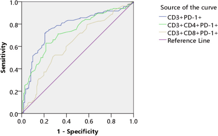
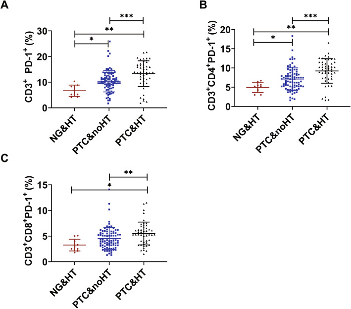
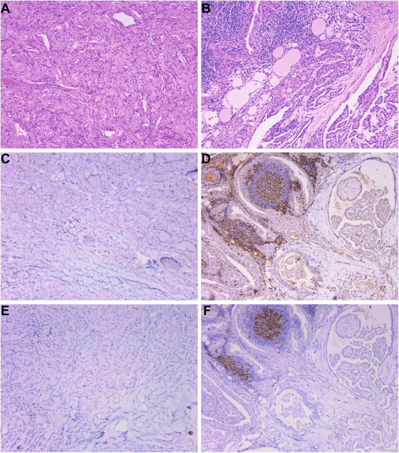
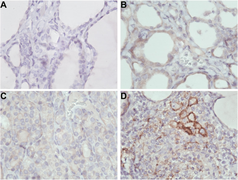
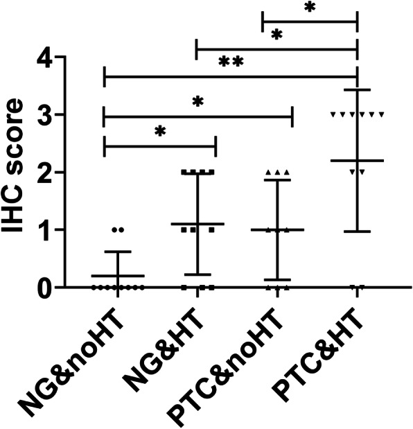
The expression of FT3, TGAb, CD3PD-1, CD3CD4PD-1 and CD3CD8PD-1 in PTC and NG was significantly higher than that in the HP group. Moreover, CD3PD-1, CD3CD4PD-1 and CD3CD8PD-1 expression had significant differences between the PTC group and the NG group. In addition, the expression of TGAb, TPO, CD3PD-1, CD3CD4PD-1 and CD3CD8PD-1 in PTC&HT group was significantly higher than that in the PTC&noHT group. While, the expression of B cells, CD3PD-1, CD3CD4PD-1 and CD3CD8PD-1 in PTC&HT group was higher than that in NG&HT group. PD-1 showed a significant correlation with PTC lymph node metastasis. CD3PD-1 and CD3CD4PD-1 was higher in N1 stage than in N0 stage. Immunohistochemical results showed that the expression of PD-1, CD3 and PD-L1 in PTC was significantly higher than that in NG.
T cell exhaustion might act as a biomarker for the differential diagnosis of PTC and NG. Patients with PTC&HT have obvious T cell exhaustion and increased expression of PD-1, PD-L1.Targeting the PD-1/PD-L1 pathway could be a new approach to prevent malignant transformation from HT to PTC&HT in the future.
在过去的几十年中,甲状腺乳头状癌(PTC)的发病率一直在稳步上升。桥本甲状腺炎(HT)是最常见的自身免疫性疾病,与 PTC 的发病机制有关。程序性死亡-1(PD-1)目前用于治疗 PTC,但关于 PD-1 在 PTC 的诊断和靶向治疗中的临床价值的研究很少。
通过流式细胞术检测 132 例 PTC 患者(PTC 组)、48 例结节性甲状腺肿(NG 组)和 63 例健康受试者(HP 组)外周血中 T、B、NK 细胞和 PD-1 的表达。采用化学发光免疫分析法检测血浆 T3、T4、FT3、FT4、TSH、TGAb 和 TPO 的表达。在 132 例 PTC 中,包括 49 例 PTC&HT 和 83 例 PTC&noHT。在 48 例 NG 中,包括 10 例 NG&HT 和 38 例 NG&noHT。通过免疫组织化学法检测 PTC 组肿瘤组织和 NG 组甲状腺组织中程序性死亡配体 1(PD-L1)、肿瘤浸润淋巴细胞(TIL)中 PD-1 和 CD3 的表达。
PTC 和 NG 组的 FT3、TGAb、CD3PD-1、CD3CD4PD-1 和 CD3CD8PD-1 表达明显高于 HP 组。此外,PTC 组与 NG 组之间 CD3PD-1、CD3CD4PD-1 和 CD3CD8PD-1 的表达差异具有统计学意义。此外,PTC&HT 组的 TGAb、TPO、CD3PD-1、CD3CD4PD-1 和 CD3CD8PD-1 表达明显高于 PTC&noHT 组。而,PTC&HT 组的 B 细胞、CD3PD-1、CD3CD4PD-1 和 CD3CD8PD-1 表达高于 NG&HT 组。PD-1 与 PTC 淋巴结转移呈显著正相关。N1 期 CD3PD-1 和 CD3CD4PD-1 高于 N0 期。免疫组化结果显示,PTC 中 PD-1、CD3 和 PD-L1 的表达明显高于 NG。
T 细胞耗竭可能作为 PTC 和 NG 鉴别诊断的生物标志物。PTC&HT 患者存在明显的 T 细胞耗竭和 PD-1、PD-L1 表达增加。靶向 PD-1/PD-L1 通路可能是预防 HT 向 PTC&HT 恶性转化的新方法。