Burch Blake, Tucker Susan
Lake Erie college of Osteopathic Medicine, Bradenton campus, Bradenton, Florida.
Radiol Case Rep. 2021 Aug 26;16(11):3182-3185. doi: 10.1016/j.radcr.2021.07.085. eCollection 2021 Nov.
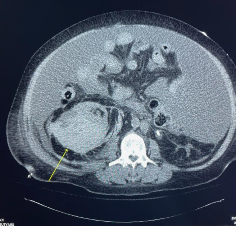
The authors report a case of a 57-year-old woman who was successfully treated with a percutaneous embolization procedure for a renal arteriovenous fistula that developed as a complication of a kidney biopsy. An acute kidney injury that failed to resolve with medical management prompted further investigation with a renal biopsy. Five hours after the kidney biopsy, the patient became hemodynamically unstable with a blood pressure of 77 of 52 mm Hg. A stat abdominal computed tomography scan without contrast discovered a large left-sided perinephric hematoma that measured up to 11.5 cm with a moderate amount of perinephric blood. An angiogram subsequently demonstrated the presence of an arteriovenous fistula at the inferior pole of the left kidney. Several 2 × 3 mm and 3 × 3 mm coils were deployed into two separate segmental branches of the inferior pole, and the post embolization angiogram confirmed resolution of the previously visualized arteriovenous fistula.
作者报告了一例57岁女性患者,该患者因肾活检并发症导致肾动静脉瘘,经皮栓塞术治疗成功。经药物治疗未能缓解的急性肾损伤促使进一步进行肾活检。肾活检5小时后,患者血流动力学不稳定,血压为77/52 mmHg。急诊非增强腹部计算机断层扫描发现左侧巨大肾周血肿,最大直径达11.5 cm,肾周有中等量血液。随后的血管造影显示左肾下极存在动静脉瘘。将几个2×3 mm和3×3 mm的线圈分别置入下极的两个节段分支,栓塞后血管造影证实先前可见的动静脉瘘已消失。