Qin Yu, Zhou Jing, Fan Zhiyuan, Gu Jianhua, Li Xinqing, Lin Dongmei, Deng Dajun, Wei Wenqiang
National Cancer Registry Office, National Cancer Center/National Clinical Research Center for Cancer/Cancer Hospital, Chinese Academy of Medical Sciences and Peking Union Medical College, Beijing, China.
Key Laboratory of Carcinogenesis and Translational Research, Peking University Cancer Hospital, Beijing, China.
Front Oncol. 2021 Aug 17;11:683876. doi: 10.3389/fonc.2021.683876. eCollection 2021.
P16 methylation is expected to be potential diagnostic and therapeutic targets for esophageal cancer (EC). The intratumoral heterogeneity (ITH) of EC has been mentioned but has not been quantitatively measured yet. We aimed to clarify the impact of ITH on pathological diagnosis and P16 methylation, and the concordance between endoscopic biopsy and the corresponding surgically resected tissue.
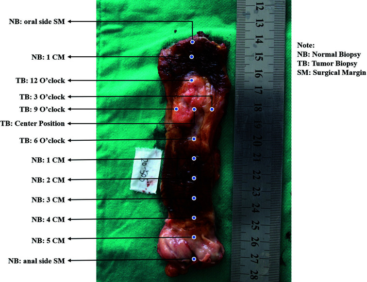
We designed a systematic sampling method (SSM) compared with a general sampling method (GSM) to obtain EC tumor tissue, tumor biopsy, and normal squamous epithelium biopsy. MethyLight assay was utilized to test P16 methylation. All specimens obtained by the SSM were pathologically diagnosed.
A total of 81 cases were collected by the GSM, and 91.4% and 8.6% of them were esophageal squamous cell carcinomas (ESCCs) and esophageal adenocarcinomas (EADs), respectively. Nine SSM cases were 100.0% ESCCs. The positive rates of P16 methylation of the GSM tumor and normal tissues were 63.0% (51/81) and 32.1% (26/81), respectively. For SSM samples, tumor tissues were 100.0% (40/40) EC and 85.0% (34/40) P16 methylated; tumor biopsy was 64.4% (29/45) diagnosed of EC and 68.9% P16 methylated; the corresponding normal biopsies were 15.7% (8/51) dysplasia and 54.9% (28/51) P16 methylated. The concordance of pathological diagnosis and P16 methylation between tumor biopsy and the corresponding tumor tissue was 75.0% and 62.5%, respectively.
The SSM we designed was efficient in measuring the ITH of EC. We found inadequate concordance between tumor biopsy and tissue in pathological diagnosis and P16 methylation.
P16甲基化有望成为食管癌(EC)潜在的诊断和治疗靶点。食管癌的肿瘤内异质性(ITH)虽已被提及,但尚未进行定量测量。我们旨在阐明ITH对病理诊断和P16甲基化的影响,以及内镜活检与相应手术切除组织之间的一致性。
我们设计了一种系统抽样方法(SSM),并与常规抽样方法(GSM)进行比较,以获取EC肿瘤组织、肿瘤活检组织和正常鳞状上皮活检组织。采用甲基化荧光定量检测法检测P16甲基化。通过SSM获取的所有标本均进行病理诊断。
GSM共收集81例病例,其中食管鳞状细胞癌(ESCC)占91.4%(74/81),食管腺癌(EAD)占8.6%(7/81)。9例SSM病例均为100.0%的ESCC。GSM肿瘤组织和正常组织中P16甲基化的阳性率分别为63.0%(51/81)和32.1%(26/81)。对于SSM样本,肿瘤组织100.0%(40/40)为EC,85.0%(34/40)P16甲基化;肿瘤活检组织64.4%(29/45)诊断为EC,68.9%P16甲基化;相应的正常活检组织15.7%(8/51)为发育异常,54.9%(28/51)P16甲基化。肿瘤活检组织与相应肿瘤组织在病理诊断和P16甲基化方面的一致性分别为75.0%和62.5%。
我们设计的SSM在测量EC的ITH方面是有效的。我们发现肿瘤活检组织与手术切除组织在病理诊断和P16甲基化方面的一致性不足。