Ion-Ebrașu Daniela, Andrei Radu Dorin, Enache Stanică, Căprărescu Simona, Negrilă Constantin Cătălin, Jianu Cătălin, Enache Adrian, Boerașu Iulian, Carcadea Elena, Varlam Mihai, Vasile Bogdan Ștefan, Ren Jianwei
National Institute for Cryogenics and Isotopic Technologies ICSI-Rm. Valcea, ICSI Energy, Uzinei Street, No. 4, 240050 Ramnicu Valcea, Romania.
Inorganic Chemistry, Physical Chemistry and Electrochemistry Department, Faculty of Applied Chemistry and Materials Science, University POLITEHNICA of Bucharest, Gh. Polizu Street, No. 1-7, 011061 Bucharest, Romania.
Materials (Basel). 2021 Aug 30;14(17):4952. doi: 10.3390/ma14174952.
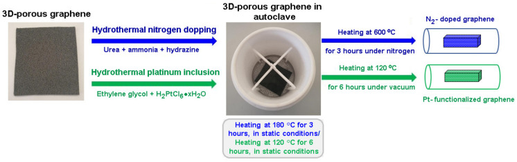
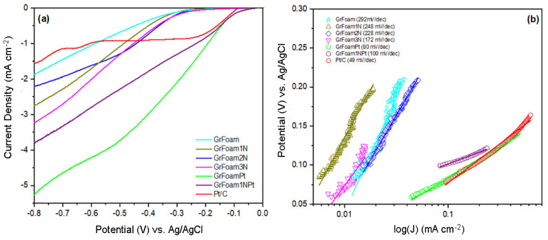
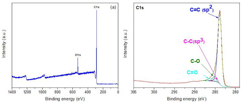
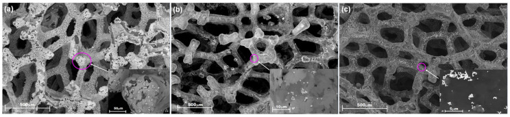
Three-dimensional graphene foam (3D-GrFoam) is a highly porous structure and sustained lattice formed by graphene layers with sp and sp hybridized carbon. In this work, chemical vapor deposition (CVD)-grown 3D-GrFoam was nitrogen-doped and platinum functionalized using hydrothermal treatment with different reducing agents (i.e., urea, hydrazine, ammonia, and dihydrogen hexachloroplatinate (IV) hydrate, respectively). X-ray photoelectron spectroscopy (XPS) survey showed that the most electrochemically active nitrogen-doped sample (GrFoam3N) contained 1.8 at % of N, and it exhibited a 172 mV dec Tafel plot associated with the Volmer-Heyrovsky hydrogen evolution (HER) mechanism in 0.1 M KOH. By the hydrothermal process, 0.2 at % of platinum was anchored to the graphene foam surface, and the resultant sample of GrFoamPt yielded a value of 80 mV dec Tafel associated with the Volmer-Tafel HER mechanism. Furthermore, Raman and infrared spectroscopy analysis, as well as scanning electron microscopy (SEM) were carried out to understand the structure of the samples.
三维石墨烯泡沫(3D-GrFoam)是一种由具有sp和sp²杂化碳的石墨烯层形成的高度多孔结构和连续晶格。在这项工作中,通过化学气相沉积(CVD)生长的3D-GrFoam使用不同的还原剂(即分别为尿素、肼、氨和六氯铂酸(IV)水合物)进行水热处理,从而实现氮掺杂和铂功能化。X射线光电子能谱(XPS)调查显示,电化学活性最高的氮掺杂样品(GrFoam3N)含氮量为1.8原子%,并且在0.1 M KOH中,其与Volmer-Heyrovsky析氢(HER)机制相关的塔菲尔斜率为172 mV dec⁻¹。通过水热过程,0.2原子%的铂锚定在石墨烯泡沫表面,所得的GrFoamPt样品的塔菲尔值为80 mV dec⁻¹,与Volmer-Tafel析氢机制相关。此外,还进行了拉曼光谱和红外光谱分析以及扫描电子显微镜(SEM)分析,以了解样品的结构。