He Jianli, Xuan Xianjun, Jiang Minhai, Li Jiangtao, Li Ning, Nie Tian
Department of Neurology, Affiliated Hangzhou First People's Hospital, Zhejiang University School of Medicine, Hangzhou, Zhejiang 310006, P.R. China.
The Fourth Clinical College, Zhejiang Chinese Medical University, Hangzhou, Zhejiang 310000, P.R. China.
Exp Ther Med. 2021 Oct;22(4):1148. doi: 10.3892/etm.2021.10581. Epub 2021 Aug 9.
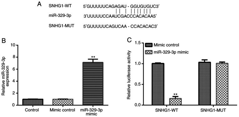
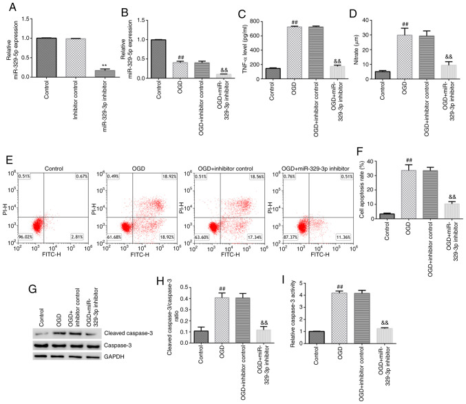
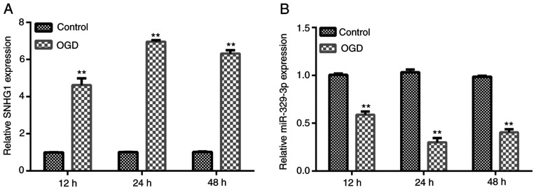
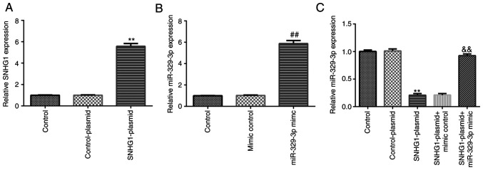
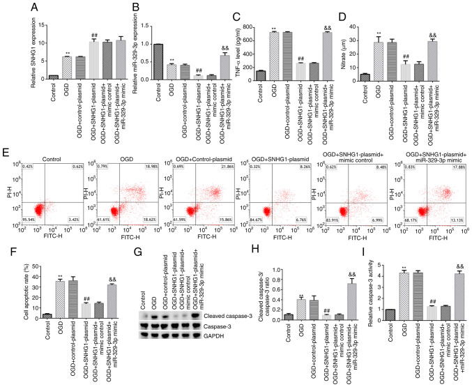
Following cerebral infarction, activated microglia cells can release a large amount of inflammatory cytokines, thereby exacerbating neuronal damage. It has been demonstrated that the long non-coding RNA small nucleolar RNA host gene 1 (SNHG1) exerts a protective effect against cerebral infarction. However, its specific role in cerebral infarction and underlying mechanism have yet to be fully elucidated. The present study aimed to investigate the effects of the SNHG1 and microRNA (miR)-329-3p in cerebral infarction and to determine the underlying molecular mechanisms. An oxygen-glucose deprivation (OGD) model was established using the BV-2 microglial cell line. The mRNA expression levels of SNHG1 and miR-329-3p were analyzed using reverse transcription-quantitative PCR and the protein expression levels of cleaved caspase-3 and caspase-3 were detected using western blotting. The binding relationship between SNHG1 and miR-329-3p was predicted using starBase and verified using a dual luciferase reporter assay. The release of TNF-α and nitric oxide, as well as caspase-3 activity, were detected using appropriate commercial kits. Flow cytometry analysis was performed to measure cell apoptosis. The results of the present study revealed that the expression levels of SNHG1 were upregulated in the OGD-induced BV-2 cell model. miR-329-3p was discovered to directly target SNHG1, and its mRNA expression levels were downregulated in the OGD-induced BV-2 cell model. The SNHG1-plasmid downregulated miR-329-3p expression levels, while this effect was reversed by transfection with the miR-329-3p mimic. The overexpression of SNHG1 or knockdown of miR-329-3p inhibited OGD-induced BV-2 cell activation. In conclusion, the results of the present study suggested that SNHG1 may reduce microglial cell activity by regulating the expression of miR-329-3p, indicating its potential protective role in cerebral infarction.
脑梗死发生后,活化的小胶质细胞可释放大量炎性细胞因子,从而加重神经元损伤。已有研究表明,长链非编码RNA小核仁RNA宿主基因1(SNHG1)对脑梗死具有保护作用。然而,其在脑梗死中的具体作用及潜在机制尚未完全阐明。本研究旨在探讨SNHG1和微小RNA(miR)-329-3p在脑梗死中的作用,并确定其潜在的分子机制。利用BV-2小胶质细胞系建立氧糖剥夺(OGD)模型。采用逆转录定量PCR分析SNHG1和miR-329-3p的mRNA表达水平,采用蛋白质印迹法检测裂解的半胱天冬酶-3和半胱天冬酶-3的蛋白表达水平。利用starBase预测SNHG1与miR-329-3p的结合关系,并通过双荧光素酶报告基因实验进行验证。使用合适的商业试剂盒检测肿瘤坏死因子-α和一氧化氮的释放以及半胱天冬酶-3的活性。进行流式细胞术分析以检测细胞凋亡。本研究结果显示,在OGD诱导的BV-2细胞模型中,SNHG1的表达水平上调。发现miR-329-3p直接靶向SNHG1,且在OGD诱导的BV-2细胞模型中其mRNA表达水平下调。SNHG1质粒下调了miR-329-3p的表达水平,而转染miR-329-3p模拟物可逆转这一作用。SNHG1的过表达或miR-329-3p的敲低抑制了OGD诱导的BV-2细胞活化。总之,本研究结果表明,SNHG1可能通过调节miR-329-3p的表达来降低小胶质细胞活性,提示其在脑梗死中具有潜在的保护作用。