Zambito Giorgia, Deng Siyuan, Haeck Joost, Gaspar Natasa, Himmelreich Uwe, Censi Roberta, Löwik Clemens, Di Martino Piera, Mezzanotte Laura
Department of Radiology and Nuclear Medicine, Erasmus Medical Center, Rotterdam, Netherlands.
Department of Molecular Genetics, Erasmus Medical Center, Rotterdam, Netherlands.
Front Med (Lausanne). 2021 Aug 27;8:712367. doi: 10.3389/fmed.2021.712367. eCollection 2021.
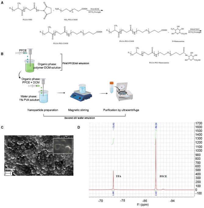
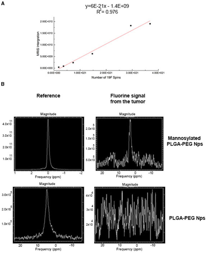
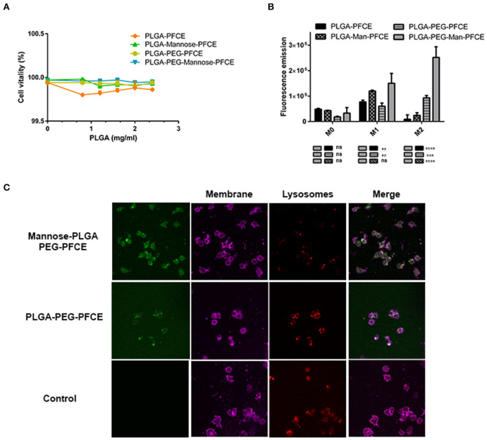
Tumor-associated macrophages (TAMs) promote cancer growth and metastasis, but their role in tumor development needs to be fully understood due to the dynamic changes of tumor microenvironment (TME). Here, we report an approach to visualize TAMs by optical imaging and by Fluorine-19 (F) magnetic resonance imaging (MRI) that is largely applied to track immune cells . TAMs are targeted with PLGA-PEG-mannose nanoparticles (NPs) encapsulating perfluoro-15-crown-5-ether (PFCE) as MRI contrast agent. These particles are preferentially recognized and phagocytized by TAMs that overexpress the mannose receptor (MRC1/CD206). The PLGA-PEG-mannose NPs are not toxic and they were up-taken by macrophages as confirmed by confocal microscopy. At 48 h after intravenous injection of PLGA-PEG-mannose NPs, 4T1 xenograft mice were imaged and fluorine-19 nuclear magnetic resonance confirmed nanoparticle retention at the tumor site. Because of the lack of F background in the body, observed F signals are robust and exhibit an excellent degree of specificity. imaging of TAMs in the TME by F MRI opens the possibility for detection of cancer at earlier stage and for prompt therapeutic interventions in solid tumors.
肿瘤相关巨噬细胞(TAMs)促进癌症生长和转移,但由于肿瘤微环境(TME)的动态变化,它们在肿瘤发展中的作用仍需充分了解。在此,我们报告了一种通过光学成像和氟-19(F)磁共振成像(MRI)可视化TAMs的方法,这种方法主要用于追踪免疫细胞。TAMs通过包裹全氟-15-冠-5-醚(PFCE)作为MRI造影剂的聚乳酸-羟基乙酸共聚物-聚乙二醇-甘露糖纳米颗粒(NPs)进行靶向。这些颗粒被过表达甘露糖受体(MRC1/CD206)的TAMs优先识别并吞噬。聚乳酸-羟基乙酸共聚物-聚乙二醇-甘露糖纳米颗粒无毒,共聚焦显微镜证实巨噬细胞摄取了这些颗粒。在静脉注射聚乳酸-羟基乙酸共聚物-聚乙二醇-甘露糖纳米颗粒48小时后,对4T1异种移植小鼠进行成像,氟-19核磁共振证实纳米颗粒在肿瘤部位滞留。由于体内缺乏F背景,观察到的F信号很强,且具有极好的特异性。通过F MRI对TME中的TAMs进行成像,为早期检测癌症以及对实体瘤进行及时的治疗干预开辟了可能性。