Division of Nephrology and Hypertension, Feinberg School of Medicine, Northwestern University, Chicago, IL, USA.
Feinberg Cardiovascular and Renal Research Institute, Feinberg School of Medicine, Northwestern University, Chicago, IL, USA.
Theranostics. 2021 Aug 27;11(18):9118-9132. doi: 10.7150/thno.60132. eCollection 2021.
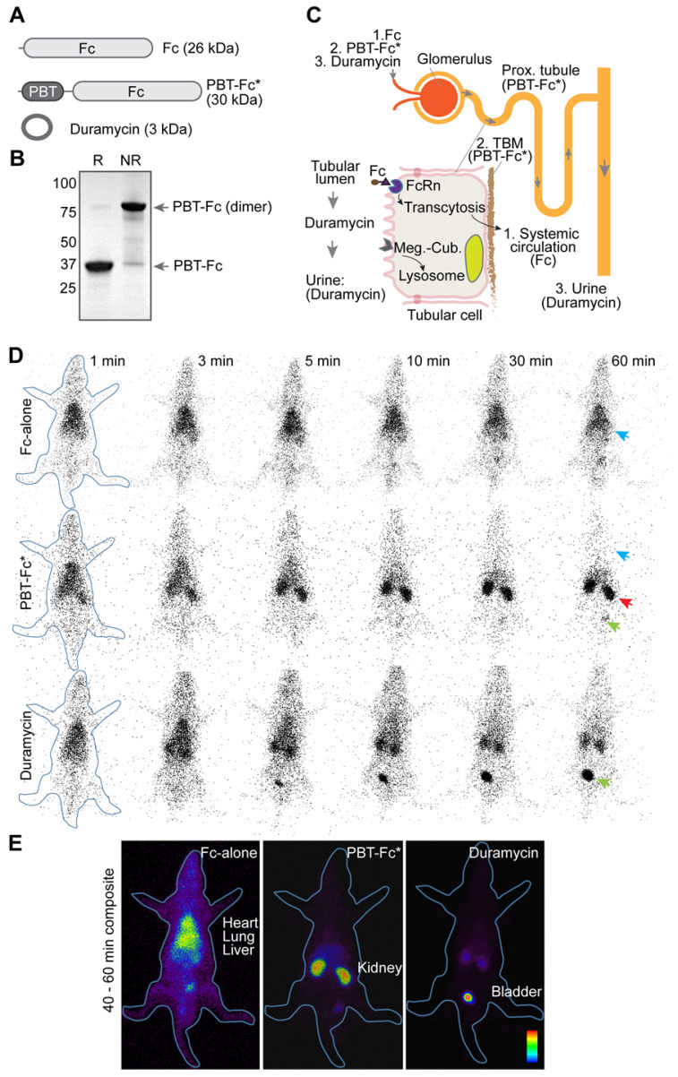
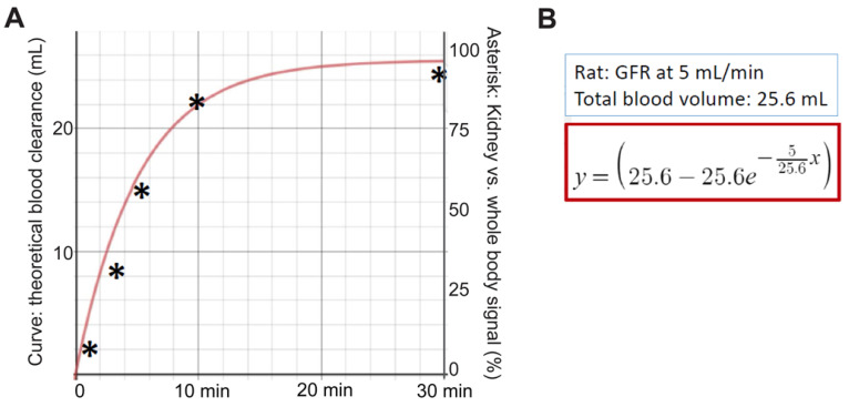
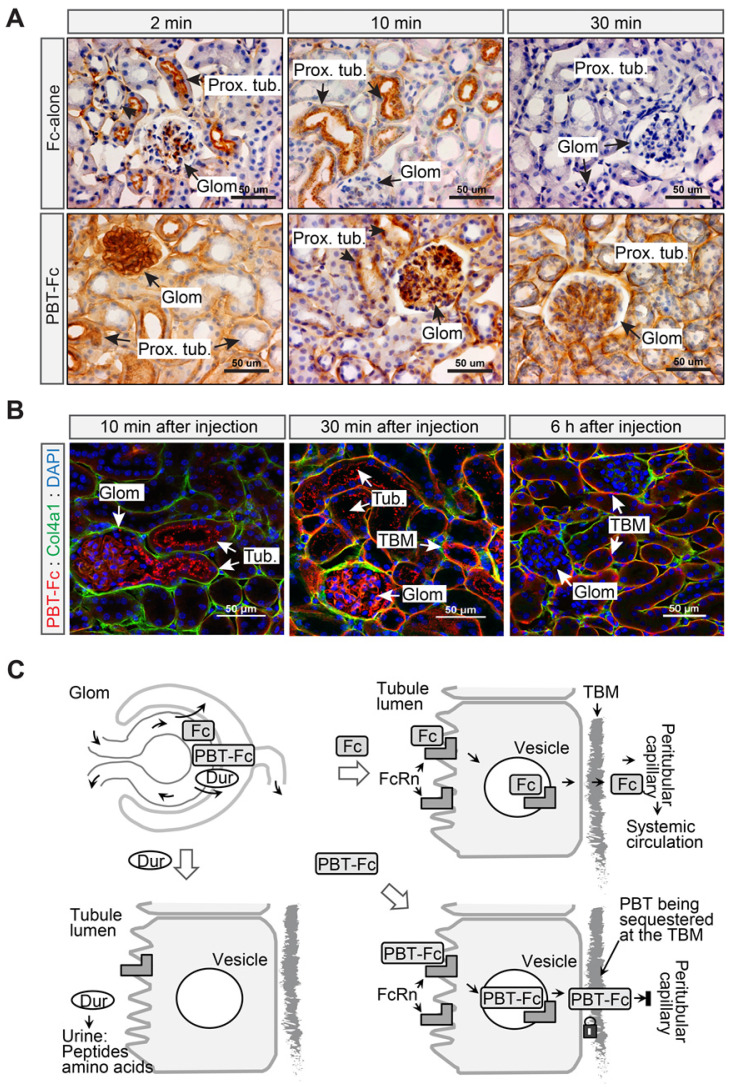
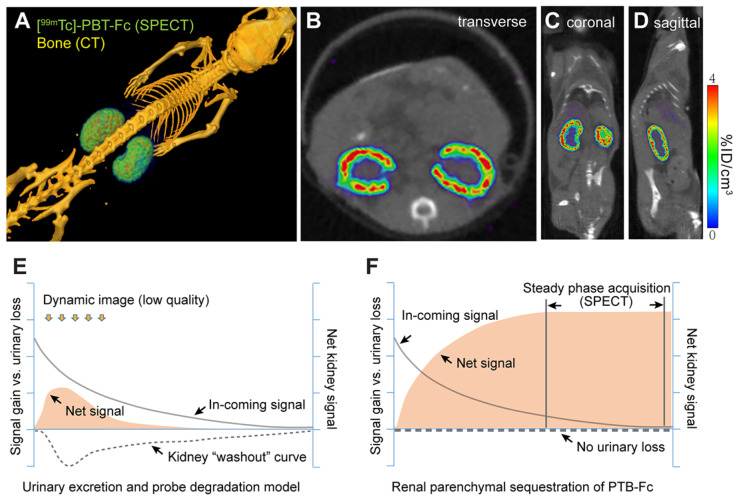
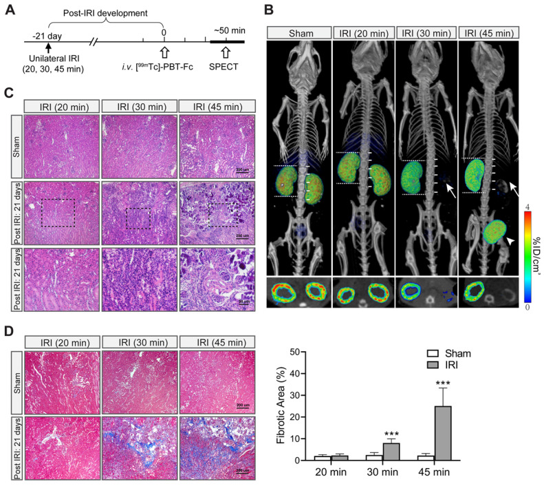
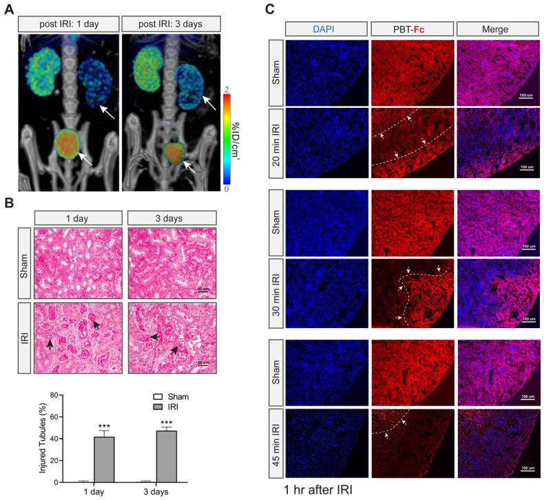
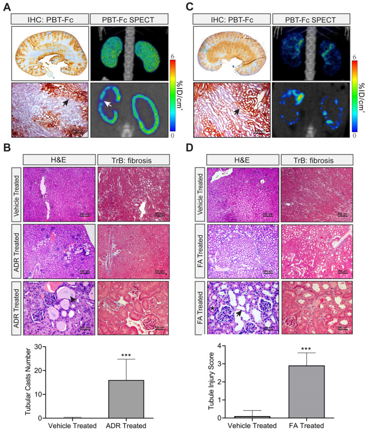
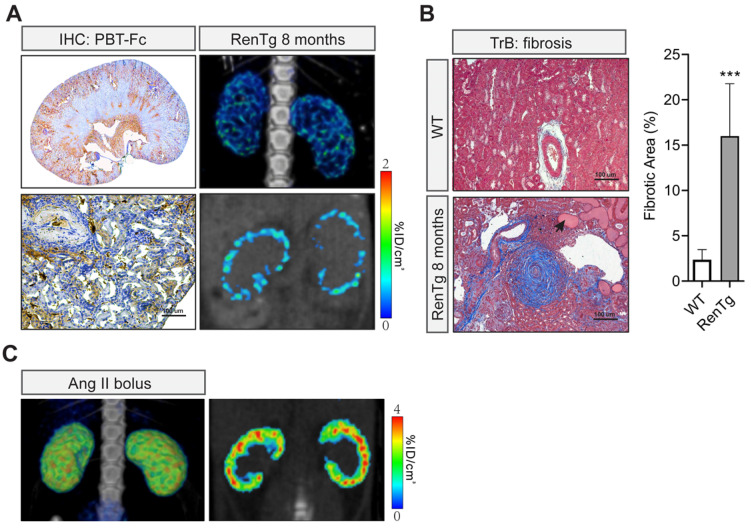
A robust radiopharmaceutical has high uptake in the target and low retention in non-target tissues. However, traditional tracers for renal imaging that chemically chelate Tc are excreted through the renal route with transient resident time in the kidney. Following a rational design approach, we constructed a protein-based radiotracer, designated PBT-Fc, to sequentially bind tubular neonatal Fc-receptor and subsequently proximal tubular basement membrane for its targeted sequestration in kidney parenchyma. In this process, the tracer participates in physiologic glomerular filtration and tubular reabsorption while escaping lysosomal catabolism and urinary clearance. To specifically target renal receptors in navigating the urinary passage in the kidney, we produced a recombinant fusion protein with two separate functional parts: a polybasic PBT segment derived from human Vascular Endothelial Growth Factor and Fc segment of IgG1. The chimeric fusion of PBT-Fc was labeled with radionuclide Tc and tested in rodent models of kidney diseases. Planar scintigraphy and single-photon emission computerized tomography (SPECT) were performed to evaluate renal-specificity of the tracer. When injected in mouse and rat, following a brief 10 - 15 min dynamic redistribution phase in circulation, ~ 95% of the [Tc]-PBT-Fc signal was concentrated in the kidney and lasted for hours without urinary loss or surrounding tissue activities. Long-lasting tracer signals in the kidney cortex in conjunction with SPECT greatly augmented the image quality in detecting pathological lesions in a variety of disease models, including ischemic acute kidney injury, drug-induced renal toxicity, and chronic kidney disease from renin-angiotensin system (RAS) overactivation. Exclusive renal retention of the recombinant radiotracer greatly facilitated static-phase signal acquisition by SPECT and achieved submillimeter spatial resolution of kidney alternations in glomerular and tubular disease models.
一种稳定的放射性药物在靶组织中有高摄取率,在非靶组织中低保留率。然而,传统的化学螯合 Tc 的肾成像示踪剂通过肾脏途径排泄,在肾脏中的停留时间短暂。通过合理的设计方法,我们构建了一种基于蛋白质的放射性示踪剂,命名为 PBT-Fc,它依次与管状新生儿 Fc 受体和随后的近端肾小管基底膜结合,以将其靶向蓄积在肾实质中。在这个过程中,示踪剂参与生理肾小球滤过和管状重吸收,同时逃避溶酶体代谢和尿液清除。为了在肾脏的尿路上专门针对肾脏受体,我们生产了一种具有两个独立功能部分的重组融合蛋白:一个来自人血管内皮生长因子的多碱性 PBT 部分和 IgG1 的 Fc 部分。PBT-Fc 的嵌合融合物用放射性核素 Tc 标记,并在肾脏疾病的啮齿动物模型中进行了测试。进行平面闪烁显像和单光子发射计算机断层扫描 (SPECT) 以评估示踪剂的肾脏特异性。当在小鼠和大鼠中注射时,在循环中的短暂 10-15 分钟的动态再分布相后,约 95%的 [Tc]-PBT-Fc 信号集中在肾脏中,并持续数小时而不会丢失尿液或周围组织活动。肾脏皮质中的长时间示踪剂信号与 SPECT 结合极大地增强了在各种疾病模型中检测病理损伤的图像质量,包括缺血性急性肾损伤、药物诱导的肾毒性和肾素-血管紧张素系统 (RAS) 过度激活引起的慢性肾病。重组放射性示踪剂的肾脏专属保留极大地促进了 SPECT 的静态相信号采集,并实现了肾小球和肾小管疾病模型中肾脏变化的亚毫米空间分辨率。