Department of Orthopaedic Surgery, Stanford University School of Medicine, 300 Pasteur Drive, Edwards R155, Stanford, CA, 94305, USA.
Department of Mechanical Engineering, Stanford University School of Medicine, Stanford, CA, USA.
Stem Cell Res Ther. 2021 Sep 15;12(1):503. doi: 10.1186/s13287-021-02572-7.
Approximately one third of patients undergoing core decompression (CD) for early-stage osteonecrosis of the femoral head (ONFH) experience progression of the disease, and subsequently require total hip arthroplasty (THA). Thus, identifying adjunctive treatments to optimize bone regeneration during CD is an unmet clinical need. Platelet-derived growth factor (PDGF)-BB plays a central role in cell growth and differentiation. The aim of this study was to characterize mesenchymal stromal cells (MSCs) that were genetically modified to overexpress PDGF-BB (PDGF-BB-MSCs) in vitro and evaluate their therapeutic effect when injected into the bone tunnel at the time of CD in an in vivo rabbit model of steroid-associated ONFH.
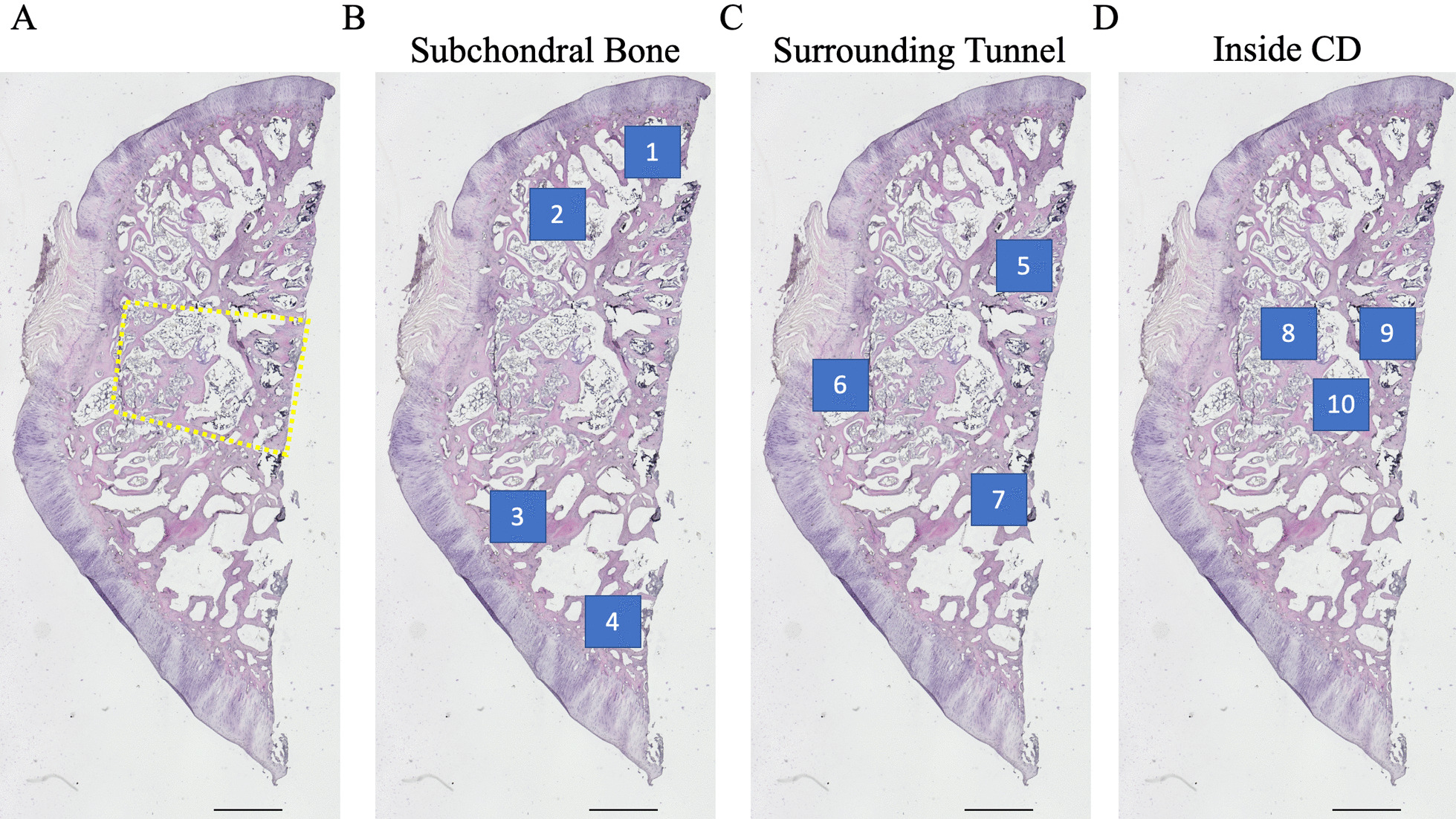
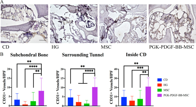
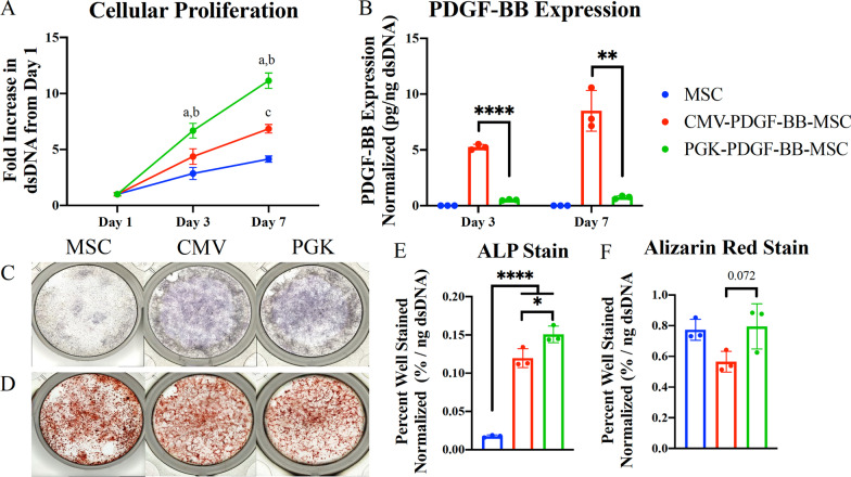
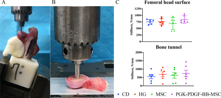
In vitro studies: Rabbit MSCs were transduced with a lentivirus vector carrying the human PDGF-BB gene under the control of either the cytomegalovirus (CMV) or phosphoglycerate (PGK) promoter. The proliferative rate, PDGF-BB expression level, and osteogenic differentiation capacity of unmodified MSCs, CMV-PDGF-BB-MSCs, and PGK-PDGF-BB-MSCs were assessed. In vivo studies: Twenty-four male New Zealand white rabbits received an intramuscular (IM) injection of methylprednisolone 20 mg/kg. Four weeks later, the rabbits were divided into four groups: the CD group, the hydrogel [HG, (a collagen-alginate mixture)] group, the MSC group, and the PGK-PDGF-BB-MSC group. Eight weeks later, the rabbits were sacrificed, their femurs were harvested, and microCT, mechanical testing, and histological analyses were performed.
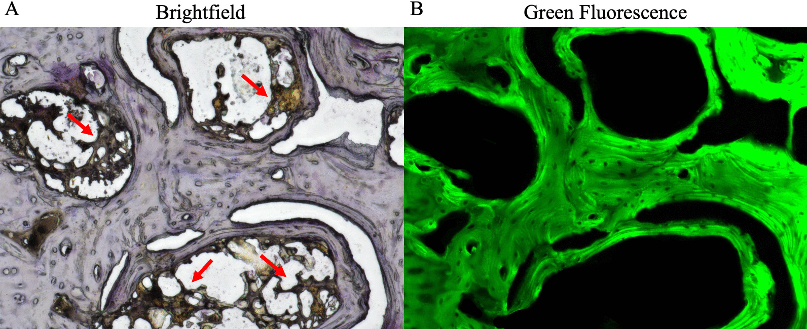
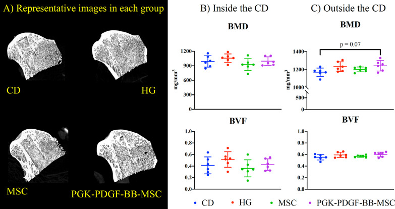
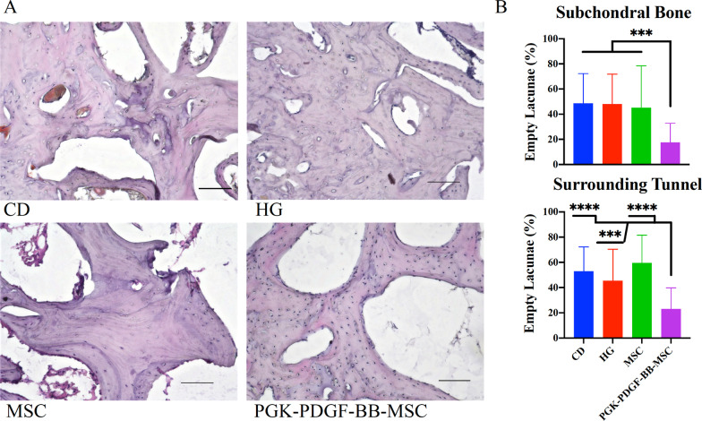
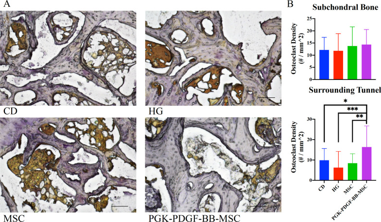
In vitro studies: PGK-PDGF-BB-MSCs proliferated more rapidly than unmodified MSCs (P < 0.001) and CMV-PDGF-BB-MSCs (P < 0.05) at days 3 and 7. CMV-PDGF-BB-MSCs demonstrated greater PDGF-BB expression than PGK-PDGF-BB-MSCs (P < 0.01). However, PGK-PDGF-BB-MSCs exhibited greater alkaline phosphatase staining at 14 days (P < 0.01), and osteogenic differentiation at 28 days (P = 0.07) than CMV-PDGF-BB-MSCs. In vivo: The PGK-PDGF-BB-MSC group had a trend towards greater bone mineral density (BMD) than the CD group (P = 0.074). The PGK-PDGF-BB-MSC group demonstrated significantly lower numbers of empty lacunae (P < 0.001), greater osteoclast density (P < 0.01), and greater angiogenesis (P < 0.01) than the other treatment groups.
The use of PGK-PDGF-BB-MSCs as an adjunctive treatment with CD may reduce progression of osteonecrosis and enhance bone regeneration and angiogenesis in the treatment of early-stage ONFH.
大约三分之一接受核心减压术 (CD) 治疗早期股骨头坏死 (ONFH) 的患者疾病会进展,随后需要全髋关节置换术 (THA)。因此,寻找辅助治疗方法以优化 CD 期间的骨再生是未满足的临床需求。血小板衍生生长因子 (PDGF)-BB 在细胞生长和分化中起核心作用。本研究的目的是对过表达 PDGF-BB 的间充质基质细胞 (MSCs) 进行体外表型鉴定,并在类固醇相关 ONFH 的兔模型中评估在 CD 时将其注射到骨隧道内的治疗效果。
体外研究:用携带人 PDGF-BB 基因的慢病毒载体转染兔 MSCs,该基因受巨细胞病毒 (CMV) 或磷酸甘油酸激酶 (PGK) 启动子的控制。评估未修饰的 MSCs、CMV-PDGF-BB-MSCs 和 PGK-PDGF-BB-MSCs 的增殖率、PDGF-BB 表达水平和成骨分化能力。体内研究:24 只雄性新西兰白兔接受肌肉内 (IM) 注射 20mg/kg 甲基强的松龙。4 周后,将兔子分为四组:CD 组、水凝胶 [HG,(胶原-海藻酸钠混合物)] 组、MSC 组和 PGK-PDGF-BB-MSC 组。8 周后,处死兔子,取出股骨,进行 microCT、力学测试和组织学分析。
体外研究:PGK-PDGF-BB-MSCs 在第 3 天和第 7 天的增殖速度比未修饰的 MSCs(P < 0.001)和 CMV-PDGF-BB-MSCs(P < 0.05)更快。CMV-PDGF-BB-MSCs 的 PDGF-BB 表达水平大于 PGK-PDGF-BB-MSCs(P < 0.01)。然而,PGK-PDGF-BB-MSCs 在第 14 天碱性磷酸酶染色(P < 0.01)和第 28 天成骨分化(P = 0.07)方面表现出更大的碱性磷酸酶染色。体内:PGK-PDGF-BB-MSC 组的骨矿物质密度 (BMD) 较 CD 组有升高趋势(P = 0.074)。PGK-PDGF-BB-MSC 组的空骨陷窝数量明显减少(P < 0.001),破骨细胞密度增加(P < 0.01),血管生成增加(P < 0.01)。
PGK-PDGF-BB-MSCs 作为 CD 的辅助治疗方法可减少骨坏死的进展,并增强早期 ONFH 治疗中的骨再生和血管生成。