Department of Neurology, North Huashan Hospital, Fudan University, No.108 Lu Xiang Road, Shanghai, 201900, China.
Department of Neurology, Huashan Hospital, Fudan University, No.12 Wulumuqi Zhong Road, Shanghai, 200040, China.
BMC Neurol. 2021 Sep 16;21(1):361. doi: 10.1186/s12883-021-02388-1.
To assess heart rate variability (HRV) among patients with arteriosclerotic cerebral small vessel disease (CSVD) by comparing with control subjects, and to determine whether HRV parameters were related to structural alterations in brain regions involved in autonomic regulation among CSVD patients.
We consecutively recruited subjects aged between 50 and 80 years who visited the Stroke Prevention Clinic of our hospital and have completed brain magnetic resonance imaging examination from September 1, 2018 to August 31, 2019. Polysomnography and synchronous analyses of HRV were then performed in all participants. Multivariable binary logistic regression was used to identify the relationship between HRV parameters and CSVD. Participants were invited to further undergo three-dimensional brain volume scan, and the voxel based morphometry (VBM) analysis was used to identify gray matter atrophy.
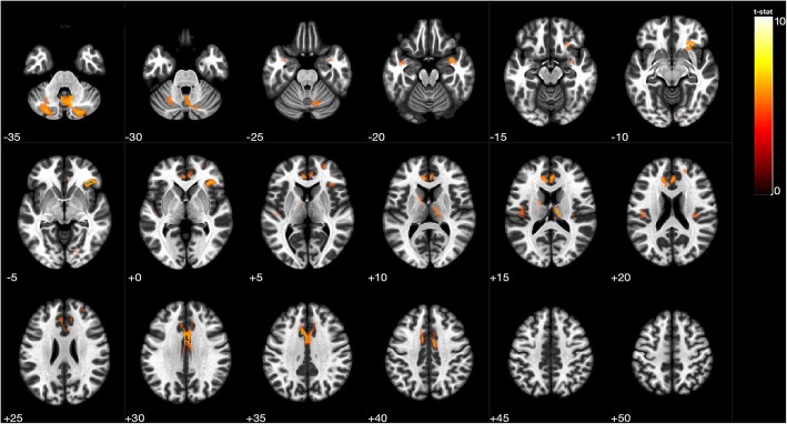
Among 109 participants enrolled in this study, 63 were assigned to the arteriosclerotic CSVD group and 46 to the control group. Lower standard deviation of normal-to-normal intervals (SDNN, OR = 0.943, 95% CI 0.903 to 0.985, P = 0.009) and higher ratio of low to high frequency power (LF/HF, OR = 4.372, 95% CI 1.033 to 18.508, P = 0.045) during the sleep period were associated with CSVD, independent of traditional cerebrovascular risk factors and sleep disordered breathing. A number of 24 CSVD patients and 21 controls further underwent three-dimensional brain volume scan and VBM analysis. Based on VBM results, SDNN during the awake time (β = 0.544, 95% CI 0.211 to 0.877, P = 0.001) and the sleep period (β = 0.532, 95% CI 0.202 to 0.862, P = 0.001) were both positively related with gray matter volume within the right inferior frontal gyrus only among CSVD patients.
Decreased nocturnal HRV is associated with arteriosclerotic CSVD independent of traditional cerebrovascular risk factors and sleep disordered breathing. The structural atrophy of some brain regions associated with cardiac autonomic regulation sheds light on the potential relationship.
Trial registration number: ChiCTR1800017902 . Date of registration: 20 Aug 2018.
通过与对照组比较,评估动脉硬化性脑小血管病(CSVD)患者的心率变异性(HRV),并确定 HRV 参数是否与 CSVD 患者自主调节相关脑区的结构改变有关。
我们连续招募了年龄在 50 至 80 岁之间的受试者,他们于 2018 年 9 月 1 日至 2019 年 8 月 31 日期间在我院卒中预防诊所就诊,并完成了脑部磁共振成像检查。然后对所有参与者进行多导睡眠图和 HRV 同步分析。采用多变量二项逻辑回归识别 HRV 参数与 CSVD 的关系。邀请参与者进一步进行三维脑容量扫描,并使用基于体素的形态测量学(VBM)分析识别灰质萎缩。
在这项研究中,共纳入 109 名参与者,其中 63 名被分配到动脉硬化性 CSVD 组,46 名被分配到对照组。睡眠期间的正常-正常间期标准差(SDNN,OR=0.943,95%CI 0.903 至 0.985,P=0.009)和低频与高频功率比(LF/HF,OR=4.372,95%CI 1.033 至 18.508,P=0.045)较低与 CSVD 相关,独立于传统的脑血管危险因素和睡眠呼吸障碍。24 名 CSVD 患者和 21 名对照组进一步进行了三维脑容量扫描和 VBM 分析。基于 VBM 结果,清醒时间(β=0.544,95%CI 0.211 至 0.877,P=0.001)和睡眠期间(β=0.532,95%CI 0.202 至 0.862,P=0.001)的 SDNN 与 CSVD 患者右侧额下回内的灰质体积呈正相关。
夜间 HRV 降低与动脉硬化性 CSVD 相关,独立于传统的脑血管危险因素和睡眠呼吸障碍。与心脏自主调节相关的一些脑区的结构萎缩提示了潜在的关系。
临床试验注册号:ChiCTR1800017902。注册日期:2018 年 8 月 20 日。