Zurich Institute of Forensic Medicine, University of Zurich, Winterthurerstrasse 190/52, 8057, Zurich, Switzerland.
Institute of Forensic Medicine, Ludwig-Maximilians-University Munich, Nussbaumstrasse 26, 80336, Munich, Germany.
Forensic Sci Med Pathol. 2021 Dec;17(4):565-576. doi: 10.1007/s12024-021-00420-x. Epub 2021 Sep 17.
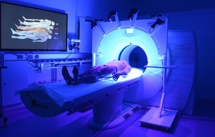
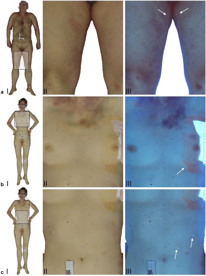
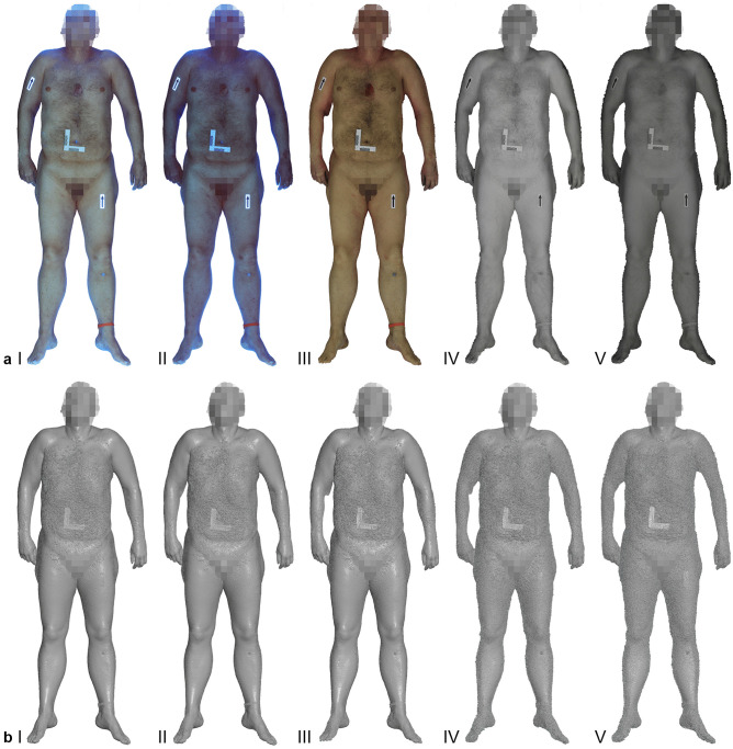
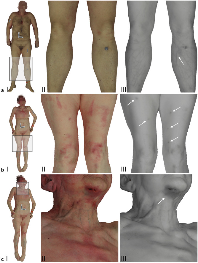
Multispectral photography offers a wide range of applications for forensic investigations. It is commonly used to detect latent evidence and to enhance the visibility of findings. Additionally, three-dimensional (3D) full-body documentation has become much easier and more affordable in recent years. However, the benefits of performing 3D imaging beyond the visible (VIS) spectrum are not well known, and the technique has not been widely used in forensic medical investigations. A multicamera setup was used to employ multispectral photogrammetry between 365 and 960 nm in postmortem investigations. The multicamera setup included four modified digital cameras, ultraviolet (UV) and near-infrared (NIR) light sources and supplemental lens filters. Full-body documentation was performed in conjunction with the use of a medical X-ray computed tomography (CT) scanner to automate the imaging procedure. Textured 3D models based on multispectral datasets from four example cases were reconstructed successfully. The level of detail and overall quality of the 3D reconstructions varied depending on the spectral range of the image data. Generally, the NIR datasets showed enhanced visibility of vein patterns and specific injuries, whereas the UV-induced datasets highlighted foreign substances on the skin. Three-dimensional multispectral full-body imaging enables the detection of latent evidence that is invisible to the naked eye and allows visualization, documentation and analysis of evidence beyond the VIS spectrum.
多光谱摄影在法医学调查中有广泛的应用。它常用于检测潜在证据并增强发现的可见度。此外,近年来,三维(3D)全身记录变得更加容易和经济实惠。然而,在可见光谱之外进行 3D 成像的好处并不为人所知,该技术在法医医学调查中尚未得到广泛应用。在法医学调查中,我们使用多相机设置在 365 至 960nm 之间进行多光谱摄影测量。该多相机设置包括四个改装的数码相机、紫外线(UV)和近红外(NIR)光源以及补充镜头滤镜。全身记录与使用医用 X 射线计算机断层扫描(CT)扫描仪相结合,以实现成像过程的自动化。成功重建了基于四个示例案例的多光谱数据集的纹理化 3D 模型。3D 重建的细节水平和整体质量因图像数据的光谱范围而异。一般来说,NIR 数据集增强了静脉模式和特定损伤的可见度,而 UV 诱导的数据集则突出了皮肤上的异物。三维多光谱全身成像能够检测肉眼不可见的潜在证据,并能够可视化、记录和分析可见光谱之外的证据。