Department of Mechanical and Industrial Engineering, University of Massachusetts, Amherst, United States.
Department of Mathematical Sciences, Worcester Polytechnic Institute, Worcester, United States.
Elife. 2021 Sep 20;10:e60381. doi: 10.7554/eLife.60381.
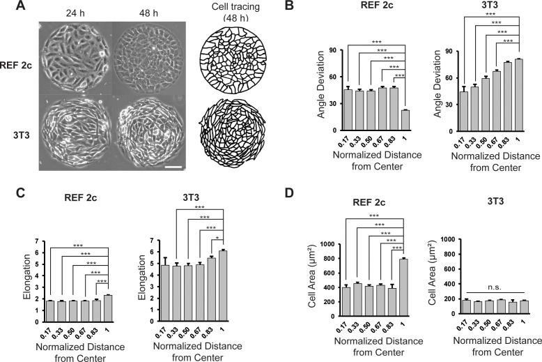
A monolayer of highly motile cells can establish long-range orientational order, which can be explained by hydrodynamic theory of active gels and fluids. However, it is less clear how cell shape changes and rearrangement are governed when the monolayer is in mechanical equilibrium states when cell motility diminishes. In this work, we report that rat embryonic fibroblasts (REF), when confined in circular mesoscale patterns on rigid substrates, can transition from the spindle shapes to more compact morphologies. Cells align radially only at the pattern boundary when they are in the mechanical equilibrium. This radial alignment disappears when cell contractility or cell-cell adhesion is reduced. Unlike monolayers of spindle-like cells such as NIH-3T3 fibroblasts with minimal intercellular interactions or epithelial cells like Madin-Darby canine kidney (MDCK) with strong cortical actin network, confined REF monolayers present an actin gradient with isotropic meshwork, suggesting the existence of a stiffness gradient. In addition, the REF cells tend to condense on soft substrates, a collective cell behavior we refer to as the 'condensation tendency'. This condensation tendency, together with geometrical confinement, induces tensile prestretch (i.e. an isotropic stretch that causes tissue to contract when released) to the confined monolayer. By developing a Voronoi-cell model, we demonstrate that the combined global tissue prestretch and cell stiffness differential between the inner and boundary cells can sufficiently define the cell radial alignment at the pattern boundary.
一层高度活跃的细胞可以建立长程取向有序,这可以用活性凝胶和流体的流体动力学理论来解释。然而,当细胞运动减弱时,单层处于力学平衡状态,细胞形状的变化和重新排列是如何被控制的,这一点就不太清楚了。在这项工作中,我们报告说,当大鼠胚胎成纤维细胞(REF)被限制在刚性基底上的圆形介观图案中时,它们可以从纺锤形转变为更紧凑的形态。当细胞处于力学平衡时,它们仅在图案边界处呈放射状排列。当细胞收缩性或细胞间粘附性降低时,这种放射状排列就会消失。与具有最小细胞间相互作用的纺锤形细胞单层(如 NIH-3T3 成纤维细胞)或具有强皮质肌动蛋白网络的上皮细胞(如 Madin-Darby 犬肾(MDCK))不同,受限的 REF 单层呈现各向同性网格的肌动蛋白梯度,表明存在刚度梯度。此外,REF 细胞往往在软基底上聚集,我们将这种集体细胞行为称为“凝聚倾向”。这种凝聚倾向与几何约束一起,对受限的单层施加拉伸预拉伸(即当释放时会导致组织收缩的各向同性拉伸)。通过开发 Voronoi 细胞模型,我们证明了组织整体预拉伸和内细胞与边界细胞之间的细胞刚度差异足以定义图案边界处的细胞放射状排列。