Nickaeen Masoud, Novak Igor L, Pulford Stephanie, Rumack Aaron, Brandon Jamie, Slepchenko Boris M, Mogilner Alex
Richard D. Berlin Center for Cell Analysis and Modeling, Department of Cell Biology, University of Connecticut Health Center, Farmington, CT, United States of America.
Center for Engineering Learning & Teaching, University of Washington, Seattle, WA, United States of America.
PLoS Comput Biol. 2017 Nov 14;13(11):e1005862. doi: 10.1371/journal.pcbi.1005862. eCollection 2017 Nov.
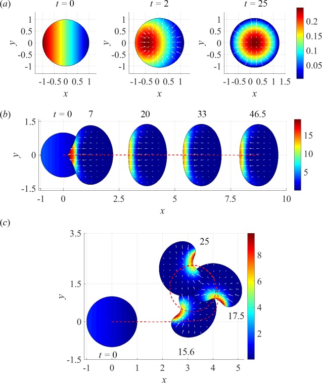
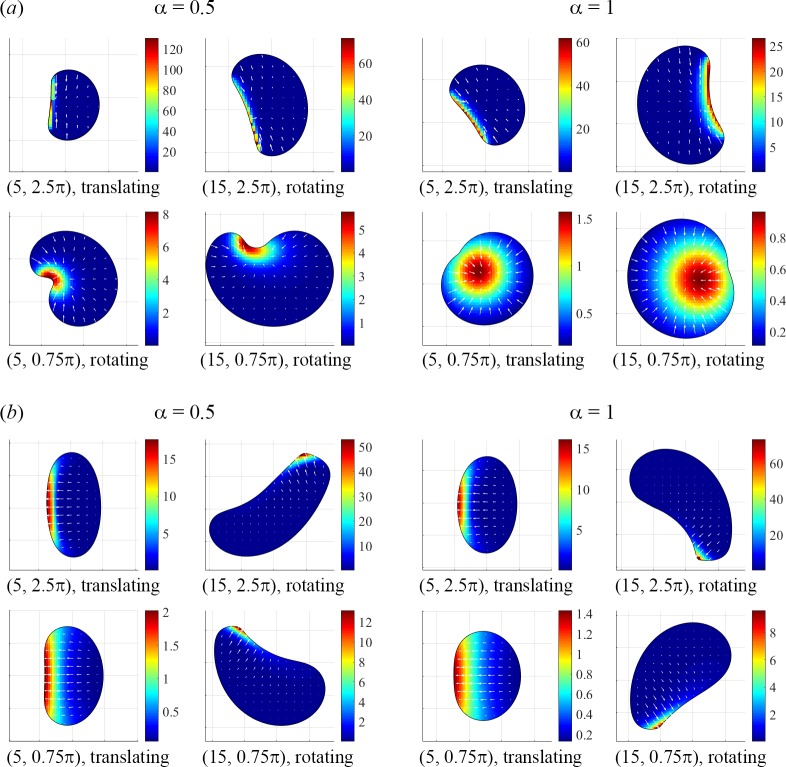
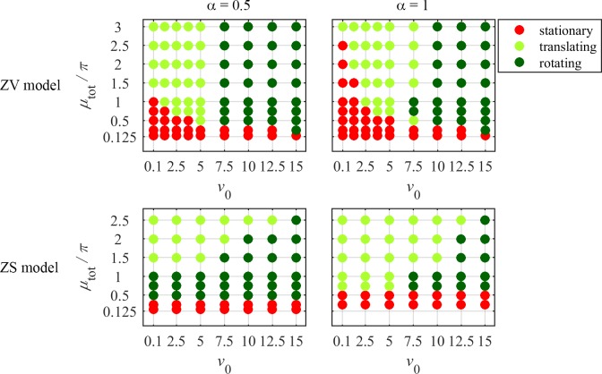
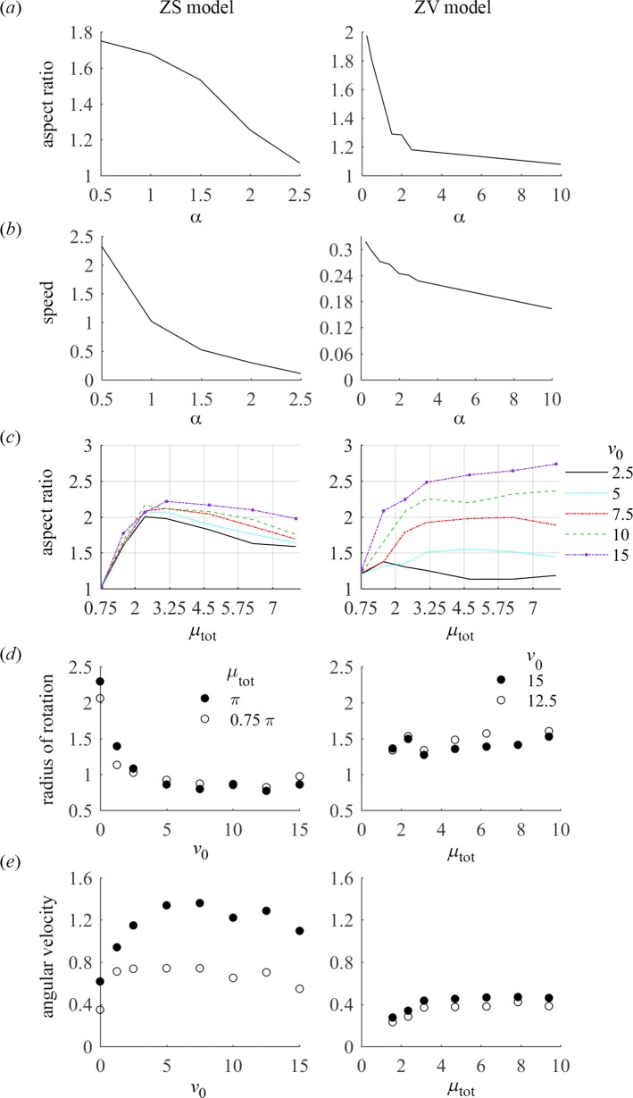
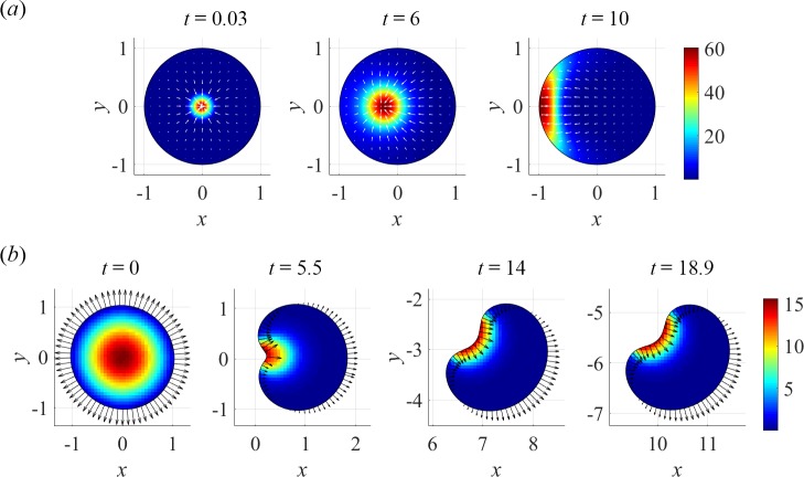
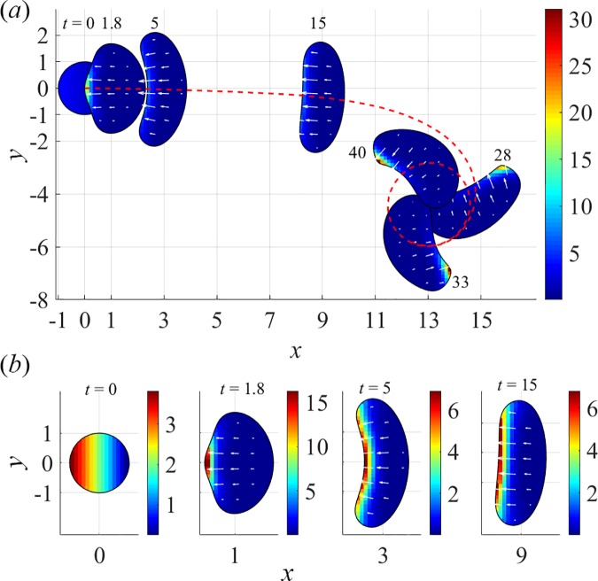
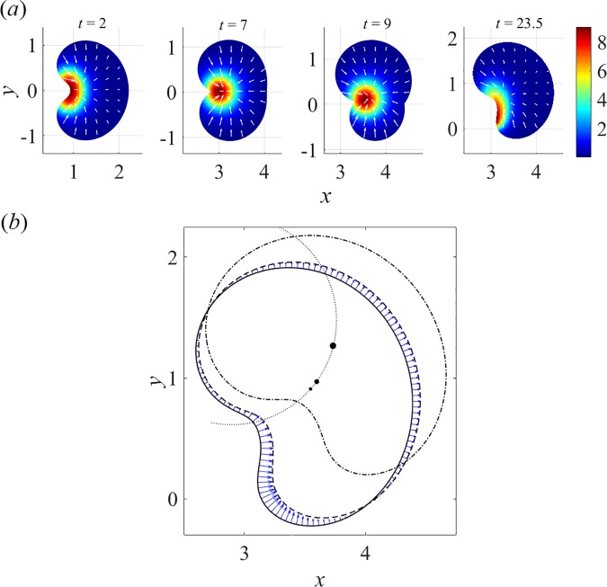
To understand shapes and movements of cells undergoing lamellipodial motility, we systematically explore minimal free-boundary models of actin-myosin contractility consisting of the force-balance and myosin transport equations. The models account for isotropic contraction proportional to myosin density, viscous stresses in the actin network, and constant-strength viscous-like adhesion. The contraction generates a spatially graded centripetal actin flow, which in turn reinforces the contraction via myosin redistribution and causes retraction of the lamellipodial boundary. Actin protrusion at the boundary counters the retraction, and the balance of the protrusion and retraction shapes the lamellipodium. The model analysis shows that initiation of motility critically depends on three dimensionless parameter combinations, which represent myosin-dependent contractility, a characteristic viscosity-adhesion length, and a rate of actin protrusion. When the contractility is sufficiently strong, cells break symmetry and move steadily along either straight or circular trajectories, and the motile behavior is sensitive to conditions at the cell boundary. Scanning of a model parameter space shows that the contractile mechanism of motility supports robust cell turning in conditions where short viscosity-adhesion lengths and fast protrusion cause an accumulation of myosin in a small region at the cell rear, destabilizing the axial symmetry of a moving cell.
为了解经历片状伪足运动的细胞的形状和运动,我们系统地探索了由力平衡和肌球蛋白传输方程组成的肌动蛋白 - 肌球蛋白收缩性的最小自由边界模型。这些模型考虑了与肌球蛋白密度成正比的各向同性收缩、肌动蛋白网络中的粘性应力以及恒定强度的类粘性粘附。收缩产生空间梯度的向心肌动蛋白流,进而通过肌球蛋白重新分布增强收缩并导致片状伪足边界的回缩。边界处的肌动蛋白突出对抗回缩,突出和回缩的平衡塑造了片状伪足的形状。模型分析表明,运动的启动关键取决于三个无量纲参数组合,它们分别代表肌球蛋白依赖性收缩性、特征粘性 - 粘附长度和肌动蛋白突出速率。当收缩性足够强时,细胞打破对称性并沿直线或圆形轨迹稳定移动,并且运动行为对细胞边界处的条件敏感。对模型参数空间的扫描表明,运动的收缩机制在短粘性 - 粘附长度和快速突出导致肌球蛋白在细胞后部的小区域积累、破坏移动细胞的轴对称性的条件下支持强大的细胞转向。