Division of Neonatology, Department of Pediatrics, Columbia University Medical Center, New York, New York, USA.
Department of Physiology & Cellular Biophysics, Columbia University, New York, New York, USA.
J Biol Chem. 2021 Oct;297(4):101204. doi: 10.1016/j.jbc.2021.101204. Epub 2021 Sep 17.
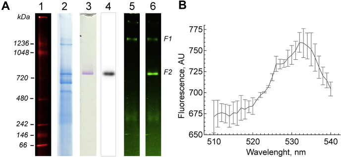
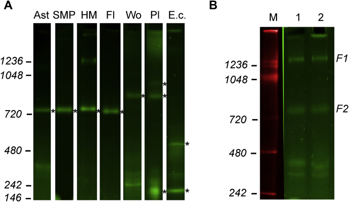
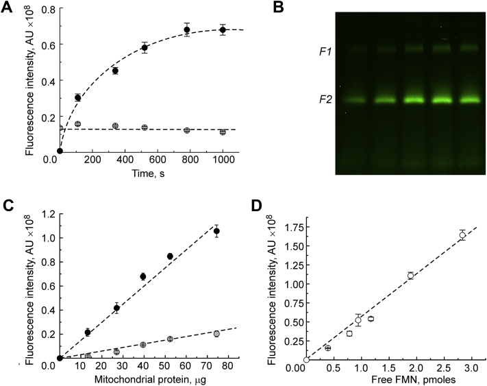
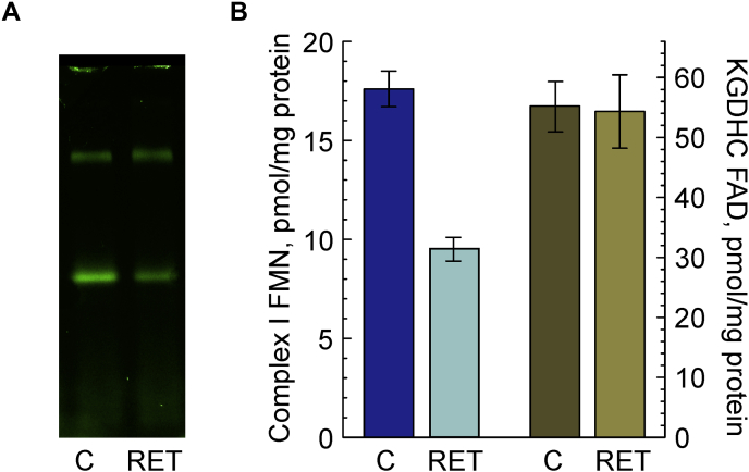
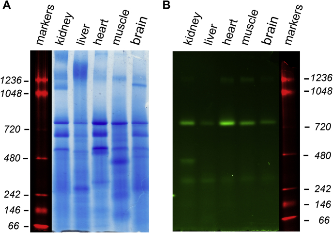
Impairments in mitochondrial energy metabolism have been implicated in human genetic diseases associated with mitochondrial and nuclear DNA mutations, neurodegenerative and cardiovascular disorders, diabetes, and aging. Alteration in mitochondrial complex I structure and activity has been shown to play a key role in Parkinson's disease and ischemia/reperfusion tissue injury, but significant difficulty remains in assessing the content of this enzyme complex in a given sample. The present study introduces a new method utilizing native polyacrylamide gel electrophoresis in combination with flavin fluorescence scanning to measure the absolute content of complex I, as well as α-ketoglutarate dehydrogenase complex, in any preparation. We show that complex I content is 19 ± 1 pmol/mg of protein in the brain mitochondria, whereas varies up to 10-fold in different mouse tissues. Together with the measurements of NADH-dependent specific activity, our method also allows accurate determination of complex I catalytic turnover, which was calculated as 10 min for NADH:ubiquinone reductase in mouse brain mitochondrial preparations. α-ketoglutarate dehydrogenase complex content was determined to be 65 ± 5 and 123 ± 9 pmol/mg protein for mouse brain and bovine heart mitochondria, respectively. Our approach can also be extended to cultured cells, and we demonstrated that about 90 × 10 complex I molecules are present in a single human embryonic kidney 293 cell. The ability to determine complex I content should provide a valuable tool to investigate the enzyme status in samples after in vivo treatment in mutant organisms, cells in culture, or human biopsies.
线粒体能量代谢的损伤与线粒体和核 DNA 突变、神经退行性和心血管疾病、糖尿病以及衰老相关的人类遗传疾病有关。已经表明,线粒体复合物 I 结构和活性的改变在帕金森病和缺血/再灌注组织损伤中起关键作用,但在评估给定样本中该酶复合物的含量方面仍存在很大困难。本研究介绍了一种新方法,利用天然聚丙烯酰胺凝胶电泳结合黄素荧光扫描来测量任何制剂中复合物 I 以及 α-酮戊二酸脱氢酶复合物的绝对含量。我们发现脑线粒体中复合物 I 的含量为 19 ± 1 pmol/mg 蛋白,而在不同的小鼠组织中变化高达 10 倍。结合 NADH 依赖性比活性的测量,我们的方法还可以准确确定复合物 I 的催化周转率,其在小鼠脑线粒体制剂中计算为 NADH:泛醌还原酶的 10 分钟。α-酮戊二酸脱氢酶复合物的含量分别为 65 ± 5 和 123 ± 9 pmol/mg 蛋白,用于小鼠脑和牛心线粒体。我们的方法也可以扩展到培养细胞,我们证明在单个人类胚胎肾 293 细胞中存在约 90×10 个复合物 I 分子。确定复合物 I 含量的能力应该为研究突变生物、培养细胞或人类活检样本中酶状态提供有价值的工具。