Department of Vascular and Endovascular Surgery, The First Affiliated Hospital of Zhengzhou University, Zhengzhou, Henan 450052, P.R. China.
Department of Hepatopancreatobiliary Surgery, The First Affiliated Hospital of Zhengzhou University, Zhengzhou, Henan 450052, P.R. China.
Oncol Rep. 2021 Nov;46(5). doi: 10.3892/or.2021.8191. Epub 2021 Sep 24.
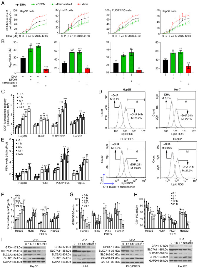
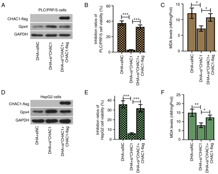
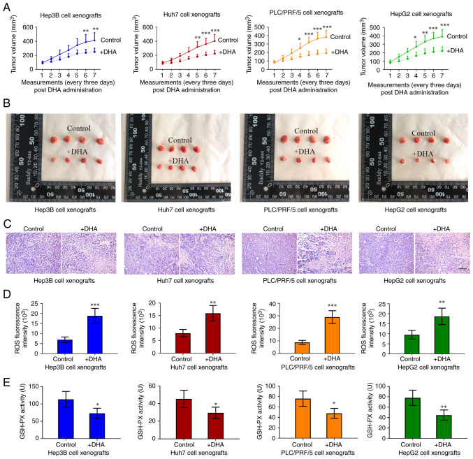
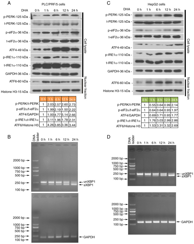
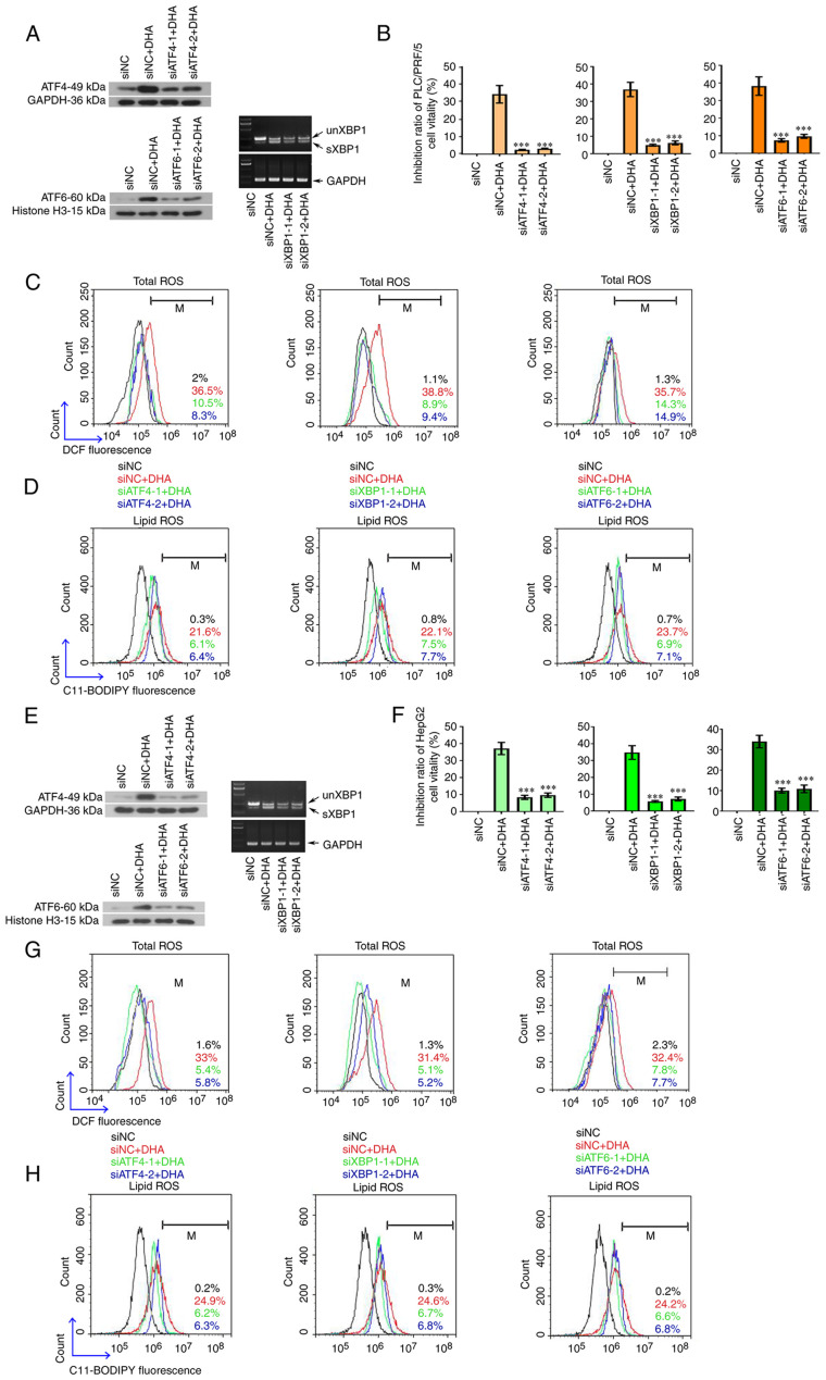
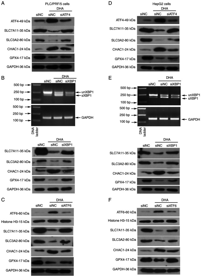
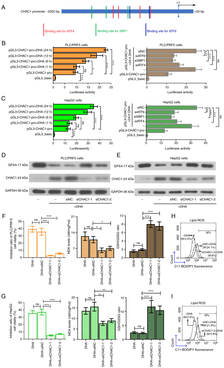
Dihydroartemisinin (DHA), an artemisinin derivate, has been investigated as a potential antitumor drug in primary liver cancer (PLC). Ferroptosis is a form of iron‑dependent cell death that can be driven by lipid peroxidation inducers. The present study aimed to determine whether and how DHA could promote the death of PLC cells by inducing ferroptosis. In total, four PLC cell lines with different p53 statuses, including Hep3B (p53 null), Huh7 (p53 mutant), PLC/PRF/5 (p53 mutant) and HepG2 (p53 wild‑type), were treated with various concentrations of DHA. The effects of DHA on all three branches of the unfolded protein response (UPR) were evaluated. To deactivate the UPRs, small interfering RNA was used to knockdown the expression of activating transcription factor (ATF)4, X‑box binding protein 1 (XBP1) or ATF6 in PLC cells. The effect of DHA on the promoter activity of Chac glutathione specific γ‑glutamylcyclotransferase 1 (CHAC1) was evaluated using a dual luciferase reporter assay. The results revealed that DHA‑induced death in PLC cells was irrelevant of the p53 status. PLC cells exposed to DHA displayed classic features of ferroptosis, such as increased lipid reactive oxygen species and malondialdehyde levels, an iron overload, and decreased activity or expression of glutathione (GSH), glutathione peroxidase 4, solute carrier family (SLC) 7 member 11 and SLC family 3 member 2. The antitumor effects of DHA in PLC cells were significantly weakened by two typical ferroptosis inhibitors, ferrostatin‑1 and deferoxamine mesylate salt, whereas the antitumor effects were augmented following iron overload. Furthermore, DHA activated all three branches of the UPR (eukaryotic translation initiation factor 2 α kinase 3/eukaryotic translation initiation factor 2A/ATF4, inositol‑requiring transmembrane kinase/endoribonuclease 1α/XBP1 and ATF6 branches) . Notably, DHA‑induced ferroptosis was significantly attenuated following the knockdown of ATF4, XBP1 or ATF6 expression. In addition, the promoter activity of CHAC1, a gene capable of degrading GSH, was enhanced by DHA, but weakened when the aforementioned three UPR transcription factors were knocked down. In conclusion, the findings of the present study suggested that DHA may effectively induce ferroptosis in PLC cells through the activation of anti‑survival UPRs and the upregulation of CHAC1 expression.
双氢青蒿素(DHA)是青蒿素的衍生物,已被研究作为原发性肝癌(PLC)的潜在抗肿瘤药物。铁死亡是一种铁依赖性细胞死亡形式,可由脂质过氧化诱导剂驱动。本研究旨在确定 DHA 是否以及如何通过诱导铁死亡来促进 PLC 细胞的死亡。总共使用了四种具有不同 p53 状态的 PLC 细胞系,包括 Hep3B(p53 缺失)、Huh7(p53 突变)、PLC/PRF/5(p53 突变)和 HepG2(p53 野生型),用不同浓度的 DHA 处理。评估了 DHA 对未折叠蛋白反应(UPR)的三条分支的影响。为了使 UPR 失活,使用小干扰 RNA 敲低 PLC 细胞中激活转录因子(ATF)4、X 框结合蛋白 1(XBP1)或 ATF6 的表达。使用双荧光素酶报告基因测定评估 DHA 对 Chac 谷胱甘肽特异性γ-谷氨酰环转移酶 1(CHAC1)启动子活性的影响。结果表明,DHA 诱导的 PLC 细胞死亡与 p53 状态无关。暴露于 DHA 的 PLC 细胞显示出铁死亡的典型特征,例如增加的脂质活性氧和丙二醛水平、铁过载以及谷胱甘肽(GSH)、谷胱甘肽过氧化物酶 4、溶质载体家族 7 成员 11 和 SLC 家族 3 成员 2 的活性或表达降低。两种典型的铁死亡抑制剂 ferrostatin-1 和甲磺酸去铁胺盐显著削弱了 DHA 在 PLC 细胞中的抗肿瘤作用,而铁过载后则增强了抗肿瘤作用。此外,DHA 激活了 UPR 的三条分支(真核翻译起始因子 2α激酶 3/真核翻译起始因子 2A/ATF4、肌醇需求跨膜激酶/内切核酸酶 1α/XBP1 和 ATF6 分支)。值得注意的是,敲低 ATF4、XBP1 或 ATF6 表达后,DHA 诱导的铁死亡明显减弱。此外,DHA 增强了能够降解 GSH 的基因 CHAC1 的启动子活性,但当敲低上述三种 UPR 转录因子时,其活性减弱。综上所述,本研究结果表明,DHA 可能通过激活抗生存 UPR 和上调 CHAC1 表达,有效诱导 PLC 细胞发生铁死亡。