Department of Radiology and Research Institute of Radiology, Asan Medical Center, University of Ulsan College of Medicine, Seoul, Korea.
Korean J Radiol. 2021 Nov;22(11):1894-1908. doi: 10.3348/kjr.2021.0248. Epub 2021 Sep 13.
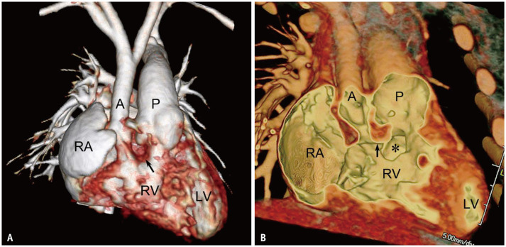
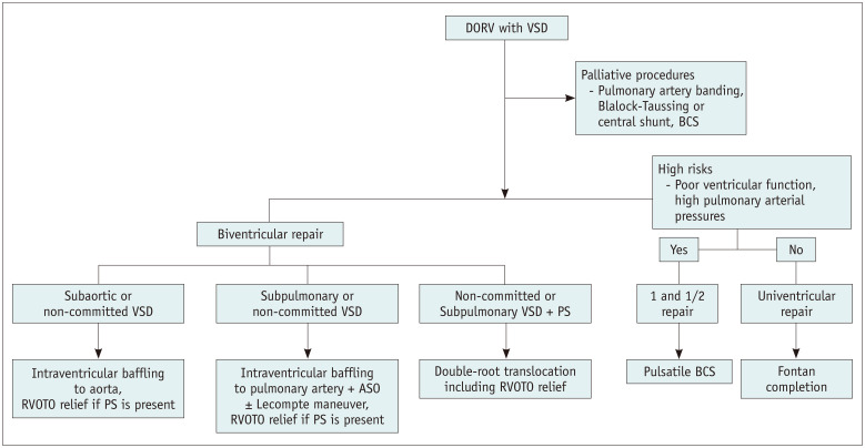
Double outlet right ventricle (DORV) is a relatively common congenital heart disease in which both great arteries are connected completely or predominantly to the morphologic RV. Unlike other congenital heart diseases, DORV demonstrates various anatomic and hemodynamic subtypes, mimicking ventricular septal defect, tetralogy of Fallot, transposition of the great arteries, and functional single ventricle. Because different surgical strategies are applied to different subtypes of DORV with ventricular septal defects, a detailed assessment of intracardiac anatomy should be performed preoperatively. Due to high spatial and contrast resolutions, cardiac CT can provide an accurate characterization of various intracardiac morphologic features of DORV. In this pictorial essay, major anatomic factors affecting surgical decision-making in DORV with ventricular septal defects were comprehensively reviewed using three-dimensional cardiac CT data. In addition, the surgical procedures available for these patients and major postoperative complications are described.
双出口右心室(DORV)是一种较为常见的先天性心脏病,两条大动脉完全或主要连接到右心室。与其他先天性心脏病不同,DORV 表现出各种解剖学和血液动力学亚型,类似于室间隔缺损、法洛四联症、大动脉转位和功能性单心室。由于不同的手术策略适用于具有室间隔缺损的不同 DORV 亚型,因此应在术前对心脏内解剖进行详细评估。由于具有较高的空间和对比分辨率,心脏 CT 可以准确描述 DORV 的各种心内形态特征。在本影像学专题论文中,使用三维心脏 CT 数据综合回顾了影响 DORV 合并室间隔缺损手术决策的主要解剖因素。此外,还描述了这些患者可采用的手术程序和主要术后并发症。