Ng Caleb C, Suresh Sandip, Rosenbaum James T, McDonald H Richard, Cunningham Emmett T
West Coast Retina Medical Group, San Francisco, CA, USA.
The Department of Ophthalmology, California Pacific Medical Center, San Francisco, CA, USA.
Am J Ophthalmol Case Rep. 2021 Sep 15;24:101206. doi: 10.1016/j.ajoc.2021.101206. eCollection 2021 Dec.
To report a series of patients with occlusive retinal vasculitis associated with systemic sclerosis (SSc) and elevated antiphospholipid antibody titers.
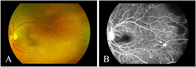
Case series. Main outcome measures included clinical and fluorescein angiographic findings at presentation and over time.
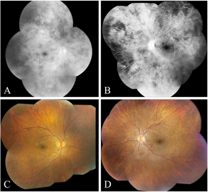
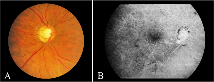
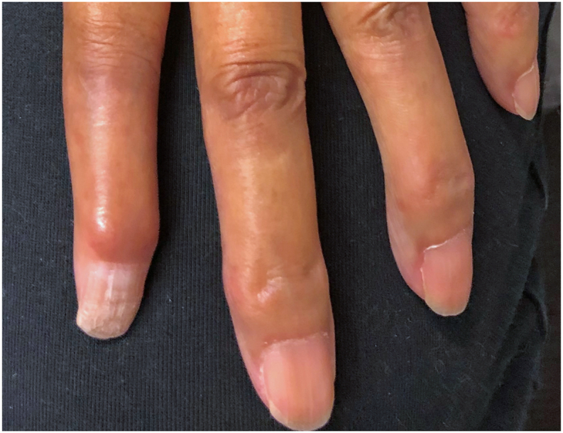
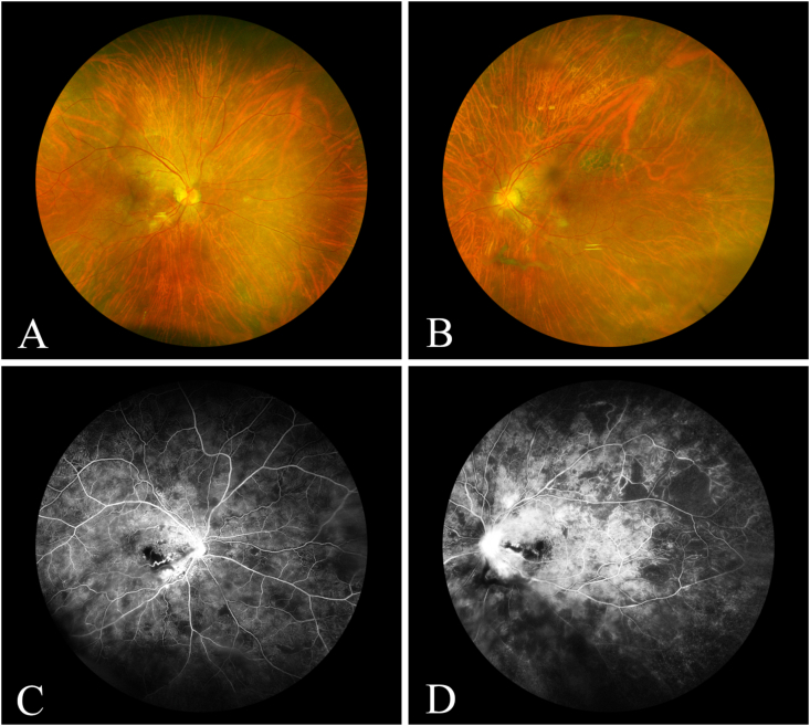
Case 1 - A 61-year-old woman initially diagnosed with idiopathic, bilateral panuveitis and retinal vasculitis causing peripheral nonperfusion was subsequently diagnosed with limited cutaneous systemic sclerosis (lcSSc). Her ocular inflammation and retinal vasculitis were controlled with topical and periocular corticosteroids, but she eventually developed peripheral retinal vascular occlusion that progressed to macular ischemia 11 years after presentation. Repeat serologic evaluation detected interval development of antiphospholipid antibodies. Case 2 - A 58-year-old woman was found to have bilateral peripheral nonperfusion and retinal neovascularization in her right eye. Given her elevated hemoglobin A1c of 8.5%, she was diagnosed with presumed proliferative diabetic retinopathy. Three years after initial presentation, she was diagnosed with lcSSc. Subsequent serum workup detected elevated B2-glycoprotein antibody titers. Her peripheral nonperfusion progressed despite adequate glycemic control, resulting in further neovascularization in each eye. Case 3 - A 40-year-old woman with diffuse cutaneous systemic sclerosis (dcSSc) and elevated titers of anti-cardiolipin antibodies developed multiple branch retinal artery occlusions with subsequent neovascularization of the retina, optic disc, and angle in the right eye.
Vision-threatening occlusive retinal vasculitis may develop in select patients with SSc. The presence of elevated anti-phospholipid antibody titers may confer increased risk for this vision-threatening complication.
报告一系列患有与系统性硬化症(SSc)相关且抗磷脂抗体滴度升高的闭塞性视网膜血管炎患者。
病例系列研究。主要观察指标包括就诊时及随访期间的临床和荧光素血管造影结果。
病例1 - 一名61岁女性最初被诊断为特发性双侧全葡萄膜炎和视网膜血管炎,导致周边视网膜无灌注,随后被诊断为局限性皮肤型系统性硬化症(lcSSc)。她的眼部炎症和视网膜血管炎通过局部和眼周皮质类固醇得到控制,但最终在就诊11年后出现周边视网膜血管闭塞,并进展为黄斑缺血。重复血清学评估发现抗磷脂抗体在随访期间出现。病例2 - 一名58岁女性右眼出现双侧周边视网膜无灌注和视网膜新生血管。鉴于其糖化血红蛋白A1c升高至8.5%,她被诊断为疑似增殖性糖尿病视网膜病变。初次就诊三年后,她被诊断为lcSSc。随后的血清学检查发现B2 - 糖蛋白抗体滴度升高。尽管血糖控制良好,她的周边视网膜无灌注仍进展,导致每只眼睛进一步出现新生血管。病例3 - 一名40岁患有弥漫性皮肤型系统性硬化症(dcSSc)且抗心磷脂抗体滴度升高的女性,右眼出现多条视网膜分支动脉闭塞,随后视网膜、视盘和房角出现新生血管。
在部分SSc患者中可能发生威胁视力的闭塞性视网膜血管炎。抗磷脂抗体滴度升高可能会增加这种威胁视力并发症的风险。