Atherosclerosis Research Unit, Department of Clinical Medicine, Aarhus University, 8200 Aarhus, Denmark.
Experimental Pathology of Atherosclerosis Laboratory, Spanish National Center for Cardiovascular Research (CNIC), 28029 Madrid, Spain.
Cells. 2021 Aug 26;10(9):2209. doi: 10.3390/cells10092209.
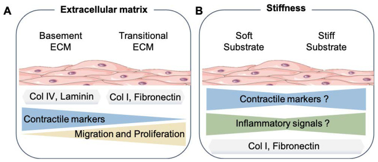
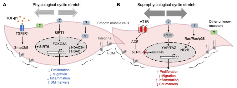
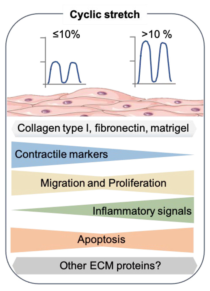
During the development of atherosclerosis and other vascular diseases, vascular smooth muscle cells (SMCs) located in the intima and media of blood vessels shift from a contractile state towards other phenotypes that differ substantially from differentiated SMCs. In addition, these cells acquire new functions, such as the production of alternative extracellular matrix (ECM) proteins and signal molecules. A similar shift in cell phenotype is observed when SMCs are removed from their native environment and placed in a culture, presumably due to the absence of the physiological signals that maintain and regulate the SMC phenotype in the vasculature. The far majority of studies describing SMC functions have been performed under standard culture conditions in which cells adhere to a rigid and static plastic plate. While these studies have contributed to discovering key molecular pathways regulating SMCs, they have a significant limitation: the ECM microenvironment and the mechanical forces transmitted through the matrix to SMCs are generally not considered. Here, we review and discuss the recent literature on how the mechanical forces and derived biochemical signals have been shown to modulate the vascular SMC phenotype and provide new perspectives about their importance.
在动脉粥样硬化和其他血管疾病的发展过程中,位于血管内膜和中膜的血管平滑肌细胞(SMC)从收缩状态向与分化的 SMC 有很大不同的其他表型转变。此外,这些细胞获得了新的功能,例如产生替代细胞外基质(ECM)蛋白和信号分子。当 SMC 从其自然环境中取出并置于培养物中时,也会观察到类似的细胞表型转变,这可能是由于缺乏维持和调节血管中 SMC 表型的生理信号。描述 SMC 功能的绝大多数研究都是在标准培养条件下进行的,其中细胞附着在刚性和静态的塑料板上。虽然这些研究有助于发现调节 SMC 的关键分子途径,但它们有一个显著的局限性:通常不考虑细胞外基质微环境和通过基质传递给 SMC 的机械力。在这里,我们回顾和讨论了最近的文献,介绍了机械力和衍生的生化信号如何被证明能够调节血管 SMC 表型,并提供了关于它们重要性的新视角。