Department of Anesthesiology and Critical Care, Medical Center-University of Freiburg, Faculty of Medicine, University of Freiburg, 79106 Freiburg, Germany.
Department of Anesthesiology, Perioperative and Pain Medicine, Brigham and Woman's Hospital, Harvard Medical School, Boston, MA 02115, USA.
Int J Mol Sci. 2021 Sep 18;22(18):10099. doi: 10.3390/ijms221810099.
The ischemia-reperfusion injury (IRI) of neuronal tissue, such as the brain and retina, leads to possible cell death and loss of function. Current treatment options are limited, but preliminary observations suggest a protective effect of hydrogen sulfide (HS). However, the dosage, timing, and mechanism of inhaled HS treatment after IRI requires further exploration.
We investigated possible neuroprotective effects of inhaled HS by inducing retinal ischemia-reperfusion injury in rats for the duration of 1 h (120 mmHg), followed by the administration of hydrogen sulfide (HS) for 1 h at different time points (0, 1.5, and 3 h after the initiation of reperfusion) and at different HS concentrations (120, 80, and 40 ppm). We quantified the HS effect by conducting retinal ganglion cell counts in fluorogold-labeled animals 7 days after IRI. The retinal tissue was harvested after 24 h for molecular analysis, including qPCR and Western blotting. Apoptotic and inflammatory mediators, transcription factors, and markers for oxidative stress were investigated. Histological analyses of the retina and the detection of inflammatory cytokines in serum assays were also performed.
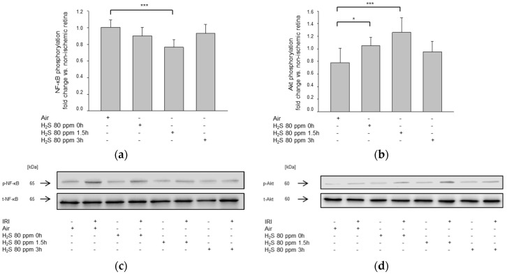
The effects of inhaled HS were most evident at a concentration of 80 ppm administered 1.5 h after IRI. HS treatment increased the expression of anti-apoptotic Bcl-2, decreased pro-apoptotic Bax expression, reduced the release of the inflammatory cytokines IL-1β and TNF-α, attenuated NF-κB p65, and enhanced Akt phosphorylation. HS also downregulated NOX4 and cystathionine β-synthase. Histological analyses illustrated a reduction in TNF-α in retinal ganglion cells and lower serum levels of TNF-α in HS-treated animals after IRI.
After neuronal IRI, HS mediates neuroprotection in a time- and dose-dependent manner. The HS treatment modulated transcription factor NF-κB activation and reduced retinal inflammation.
神经元组织(如脑和视网膜)的缺血再灌注损伤(IRI)可导致细胞死亡和功能丧失。目前的治疗选择有限,但初步观察表明硫化氢(HS)具有保护作用。然而,HS 吸入治疗在 IRI 后的剂量、时间和机制仍需进一步探索。
我们通过诱导大鼠视网膜缺血再灌注损伤 1 小时(120mmHg)来研究吸入 HS 的可能神经保护作用,随后在再灌注开始后 0、1.5 和 3 小时以及不同 HS 浓度(120、80 和 40ppm)下给予 HS 1 小时。我们通过对氟金标大鼠 7 天后的视网膜神经节细胞计数来量化 HS 的作用。在 24 小时后采集视网膜组织进行分子分析,包括 qPCR 和 Western blot。研究了凋亡和炎症介质、转录因子以及氧化应激标志物。还对视网膜进行了组织学分析,并在血清测定中检测了炎症细胞因子。
HS 浓度为 80ppm 且在 IRI 后 1.5 小时给药时,吸入 HS 的效果最为明显。HS 处理增加了抗凋亡 Bcl-2 的表达,降低了促凋亡 Bax 的表达,减少了促炎细胞因子 IL-1β 和 TNF-α的释放,减弱了 NF-κB p65,并增强了 Akt 磷酸化。HS 还下调了 NOX4 和胱硫醚-β-合酶。组织学分析表明,在 IRI 后,HS 处理降低了 TNF-α在视网膜神经节细胞中的表达,并降低了 HS 处理动物血清中 TNF-α的水平。
在神经元 IRI 后,HS 以时间和剂量依赖的方式介导神经保护作用。HS 处理调节了转录因子 NF-κB 的激活并减轻了视网膜炎症。