• 文献检索
  • 文档翻译
  • 深度研究
  • 学术资讯
  • Suppr Zotero 插件Zotero 插件
  • 邀请有礼
  • 套餐&价格
  • 历史记录
应用&插件
Suppr Zotero 插件Zotero 插件浏览器插件Mac 客户端Windows 客户端微信小程序
定价
高级版会员购买积分包购买API积分包
服务
文献检索文档翻译深度研究API 文档MCP 服务
关于我们
关于 Suppr公司介绍联系我们用户协议隐私条款
关注我们

Suppr 超能文献

核心技术专利:CN118964589B侵权必究
粤ICP备2023148730 号-1Suppr @ 2026

文献检索

告别复杂PubMed语法,用中文像聊天一样搜索,搜遍4000万医学文献。AI智能推荐,让科研检索更轻松。

立即免费搜索

文件翻译

保留排版,准确专业,支持PDF/Word/PPT等文件格式,支持 12+语言互译。

免费翻译文档

深度研究

AI帮你快速写综述,25分钟生成高质量综述,智能提取关键信息,辅助科研写作。

立即免费体验

利用原子力显微镜表征抗菌肽诱导的模型支撑脂质双层缺陷

Atomic Force Microscopy to Characterize Antimicrobial Peptide-Induced Defects in Model Supported Lipid Bilayers.

作者信息

Swana Kathleen W, Nagarajan Ramanathan, Camesano Terri A

机构信息

Department of Chemical Engineering, Worcester Polytechnic Institute, Worcester, MA 01609, USA.

U.S. Army Combat Capabilities Development Command Soldier Center, Natick, MA 01760, USA.

出版信息

Microorganisms. 2021 Sep 17;9(9):1975. doi: 10.3390/microorganisms9091975.

DOI:10.3390/microorganisms9091975
PMID:34576869
原文链接:https://pmc.ncbi.nlm.nih.gov/articles/PMC8465339/
Abstract

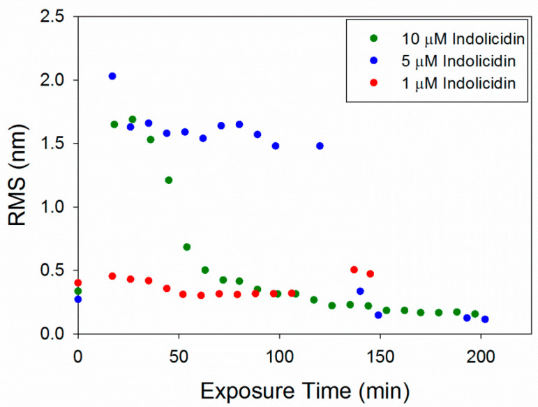
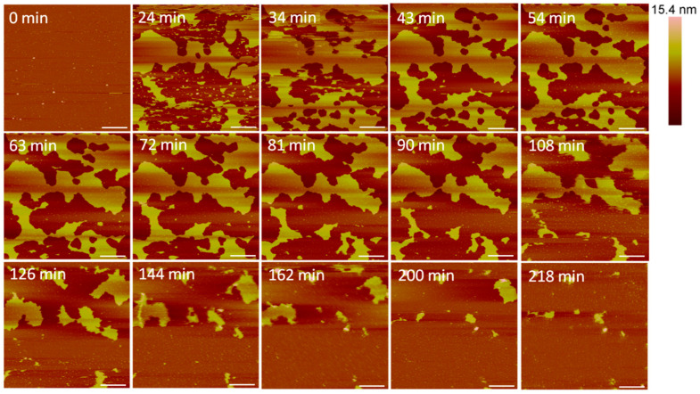
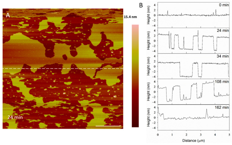
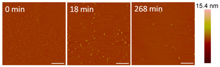
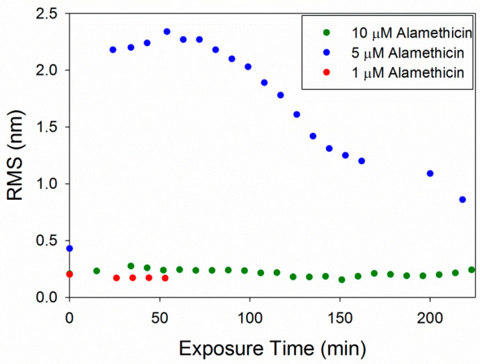
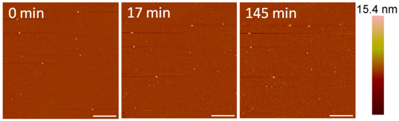
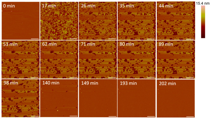
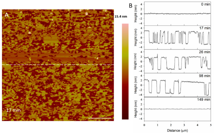
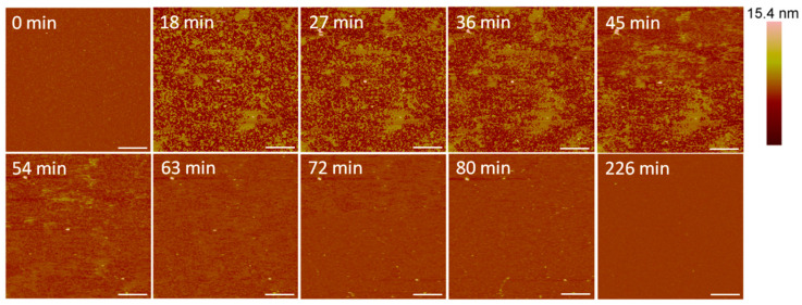
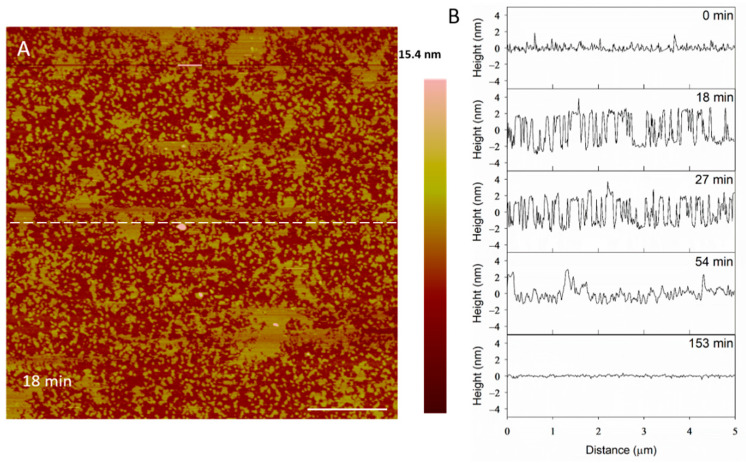
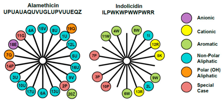
Antimicrobial peptides (AMPs) interact with bacterial cell membranes through a variety of mechanisms, causing changes extending from nanopore formation to microscale membrane lysis, eventually leading to cell death. Several AMPs also disrupt mammalian cell membranes, despite their significantly different lipid composition and such collateral hemolytic damage hinders the potential therapeutic applicability of the AMP as an anti-microbial. Elucidating the mechanisms underlying the AMP-membrane interactions is challenging due to the variations in the chemical and structural features of the AMPs, the complex compositional variations of cell membranes and the inadequacy of any single experimental technique to comprehensively probe them. (1) Background: Atomic Force Microscopy (AFM) imaging can be used in combination with other techniques to help understand how AMPs alter the orientation and structural organization of the molecules within cell membranes exposed to AMPs. The structure, size, net charge, hydrophobicity and amphipathicity of the AMPs affect how they interact with cell membranes of differing lipid compositions. (2) Methods: Our study examined two different types of AMPs, a 20-amino acid, neutral, α-helical (amphipathic) peptide, alamethicin, and a 13-amino acid, non-α-helical cationic peptide, indolicidin (which intramolecularly folds, creating a hydrophobic core), for their interactions with supported lipid bilayers (SLBs). Robust SLB model membranes on quartz supports, incorporating predominantly anionic lipids representative of bacterial cells, are currently not available and remain to be developed. Therefore, the SLBs of zwitterionic egg phosphatidylcholine (PC), which represents the composition of a mammalian cell membrane, was utilized as the model membrane. This also allows for a comparison with the results obtained from the Quartz Crystal Microbalance with Dissipation (QCM-D) experiments conducted for these peptides interacting with the same zwitterionic SLBs. Further, in the case of alamethicin, because of its neutrality, the lipid charge may be less relevant for understanding its membrane interactions. (3) Results: Using AFM imaging and roughness analysis, we found that alamethicin produced large, unstable defects in the membrane at 5 µM concentrations, and completely removed the bilayer at 10 µM. Indolicidin produced smaller holes in the bilayer at 5 and 10 µM, although they were able to fill in over time. The root-mean-square (RMS) roughness values for the images showed that the surface roughness caused by visible defects peaked after peptide injection and gradually decreased over time. (4) Conclusions: AFM is useful for helping to uncover the dynamic interactions between different AMPs and cell membranes, which can facilitate the selection and design of more efficient AMPs for use in therapeutics and antimicrobial applications.

https://cdn.ncbi.nlm.nih.gov/pmc/blobs/e943/8465339/255a71f32394/microorganisms-09-01975-g012.jpg
https://cdn.ncbi.nlm.nih.gov/pmc/blobs/e943/8465339/8f41696273d3/microorganisms-09-01975-g001.jpg
https://cdn.ncbi.nlm.nih.gov/pmc/blobs/e943/8465339/8619641c1169/microorganisms-09-01975-g002.jpg
https://cdn.ncbi.nlm.nih.gov/pmc/blobs/e943/8465339/5d09b584a959/microorganisms-09-01975-g003.jpg
https://cdn.ncbi.nlm.nih.gov/pmc/blobs/e943/8465339/43a56c95726f/microorganisms-09-01975-g004.jpg
https://cdn.ncbi.nlm.nih.gov/pmc/blobs/e943/8465339/21df5f435d5a/microorganisms-09-01975-g005.jpg
https://cdn.ncbi.nlm.nih.gov/pmc/blobs/e943/8465339/a0903067fc5f/microorganisms-09-01975-g006.jpg
https://cdn.ncbi.nlm.nih.gov/pmc/blobs/e943/8465339/5d0a8a18db7d/microorganisms-09-01975-g007.jpg
https://cdn.ncbi.nlm.nih.gov/pmc/blobs/e943/8465339/4207c90622e1/microorganisms-09-01975-g008.jpg
https://cdn.ncbi.nlm.nih.gov/pmc/blobs/e943/8465339/eb107378a630/microorganisms-09-01975-g009.jpg
https://cdn.ncbi.nlm.nih.gov/pmc/blobs/e943/8465339/8fca8886a39b/microorganisms-09-01975-g010.jpg
https://cdn.ncbi.nlm.nih.gov/pmc/blobs/e943/8465339/08b30112b665/microorganisms-09-01975-g011.jpg
https://cdn.ncbi.nlm.nih.gov/pmc/blobs/e943/8465339/255a71f32394/microorganisms-09-01975-g012.jpg
摘要

抗菌肽(AMPs)通过多种机制与细菌细胞膜相互作用,引起从纳米孔形成到微观尺度膜裂解等一系列变化,最终导致细胞死亡。尽管哺乳动物细胞膜的脂质组成与细菌细胞膜显著不同,但仍有几种抗菌肽会破坏其细胞膜,这种附带的溶血损伤阻碍了抗菌肽作为抗菌剂的潜在治疗应用。由于抗菌肽的化学和结构特征存在差异、细胞膜的复杂组成变化以及任何单一实验技术都不足以全面探测这些相互作用,因此阐明抗菌肽与膜相互作用的潜在机制具有挑战性。(1)背景:原子力显微镜(AFM)成像可与其他技术结合使用,以帮助理解抗菌肽如何改变暴露于抗菌肽的细胞膜内分子的取向和结构组织。抗菌肽的结构、大小、净电荷、疏水性和两亲性会影响它们与不同脂质组成的细胞膜的相互作用方式。(2)方法:我们的研究考察了两种不同类型的抗菌肽,一种是由20个氨基酸组成的中性α-螺旋(两亲性)肽阿拉米辛,另一种是由13个氨基酸组成的非α-螺旋阳离子肽吲哚杀菌素(其分子内折叠形成疏水核心),研究它们与支持脂质双层(SLBs)的相互作用。目前尚无包含主要代表细菌细胞的阴离子脂质的、在石英载体上的稳健的支持脂质双层模型膜,仍有待开发。因此,两性离子型卵磷脂(PC)的支持脂质双层被用作模型膜,其代表哺乳动物细胞膜的组成。这也便于与这些肽与相同两性离子支持脂质双层相互作用的石英晶体微天平耗散技术(QCM-D)实验结果进行比较。此外,对于阿拉米辛,由于其呈中性,脂质电荷对于理解其与膜的相互作用可能不太相关。(3)结果:通过AFM成像和粗糙度分析,我们发现5μM浓度的阿拉米辛会在膜中产生大的、不稳定的缺陷,而在10μM时会完全破坏双层膜。5μM和10μM的吲哚杀菌素会在双层膜中产生较小的孔,不过随着时间推移这些孔能够被填充。图像的均方根(RMS)粗糙度值表明,由可见缺陷引起的表面粗糙度在注入肽后达到峰值,并随时间逐渐降低。(4)结论:AFM有助于揭示不同抗菌肽与细胞膜之间的动态相互作用,这有助于选择和设计更有效的抗菌肽用于治疗和抗菌应用。

相似文献

1
Atomic Force Microscopy to Characterize Antimicrobial Peptide-Induced Defects in Model Supported Lipid Bilayers.利用原子力显微镜表征抗菌肽诱导的模型支撑脂质双层缺陷
Microorganisms. 2021 Sep 17;9(9):1975. doi: 10.3390/microorganisms9091975.
2
Formation of a Fully Anionic Supported Lipid Bilayer to Model Bacterial Inner Membrane for QCM-D Studies.形成完全阴离子支撑脂质双层以模拟用于石英晶体微天平耗散监测研究的细菌内膜。
Membranes (Basel). 2022 May 27;12(6):558. doi: 10.3390/membranes12060558.
3
Differentiating antimicrobial peptides interacting with lipid bilayer: Molecular signatures derived from quartz crystal microbalance with dissipation monitoring.区分与脂质双层相互作用的抗菌肽:源自具有耗散监测功能的石英晶体微天平的分子特征。
Biophys Chem. 2015 Jan;196:53-67. doi: 10.1016/j.bpc.2014.09.003. Epub 2014 Sep 28.
4
Antimicrobial peptide alamethicin insertion into lipid bilayer: a QCM-D exploration.抗菌肽阿拉米辛插入脂质双层:石英晶体微天平耗散技术(QCM-D)研究
Colloids Surf B Biointerfaces. 2014 Apr 1;116:472-81. doi: 10.1016/j.colsurfb.2014.01.036. Epub 2014 Jan 31.
5
Mechanistic predictions of the influence of collagen-binding domain sequences on human LL37 interactions with model lipids using quartz crystal microbalance with dissipation.利用石英晶体微天平的耗散技术对胶原蛋白结合域序列对人 LL37 与模型脂质相互作用的影响进行机理预测。
Biointerphases. 2019 Apr 30;14(2):021006. doi: 10.1116/1.5089759.
6
Role of lipopolysaccharides and lipoteichoic acids on C-Chrysophsin-1 interactions with model Gram-positive and Gram-negative bacterial membranes.脂多糖和脂磷壁酸在 C-Chrysophsin-1 与模型革兰氏阳性和革兰氏阴性细菌膜相互作用中的作用。
Biointerphases. 2020 May 26;15(3):031007. doi: 10.1116/1.5130774.
7
Characterisation of cell membrane interaction mechanisms of antimicrobial peptides by electrical bilayer recording.用电双层记录技术研究抗菌肽与细胞膜相互作用机制的特性
Biophys Chem. 2022 Feb;281:106721. doi: 10.1016/j.bpc.2021.106721. Epub 2021 Nov 16.
8
The potential of AFM in studying the role of the nanoscale amphipathic nature of (lipo)-peptides interacting with lipid bilayers.原子力显微镜在研究(脂)肽的纳米级两亲性与脂质双层相互作用中的作用的潜力。
Nanotechnology. 2022 Aug 1;33(43). doi: 10.1088/1361-6528/ac80c9.
9
Cationic peptide-induced remodelling of model membranes: direct visualization by in situ atomic force microscopy.阳离子肽诱导的模型膜重塑:原位原子力显微镜直接可视化
J Struct Biol. 2008 Apr;162(1):121-38. doi: 10.1016/j.jsb.2007.11.003. Epub 2007 Nov 17.
10
A biophysical study of the interactions between the antimicrobial peptide indolicidin and lipid model systems.抗菌肽吲哚西林与类脂模型系统相互作用的生物物理研究。
Biochim Biophys Acta Biomembr. 2019 Jul 1;1861(7):1355-1364. doi: 10.1016/j.bbamem.2019.04.003. Epub 2019 Apr 9.

引用本文的文献

1
Boosting AMPs' Power: From Structural Engineering to Nanotechnology-Based Delivery.增强抗菌肽的功效:从结构工程到基于纳米技术的递送
Molecules. 2025 Jul 15;30(14):2979. doi: 10.3390/molecules30142979.
2
Short-Chained Linear Scorpion Peptides: A Pool for Novel Antimicrobials.短链线性蝎肽:新型抗菌剂的宝库
Antibiotics (Basel). 2024 May 5;13(5):422. doi: 10.3390/antibiotics13050422.
3
Effect of CM15 on Supported Lipid Bilayer Probed by Atomic Force Microscopy.原子力显微镜探测CM15对支撑脂质双层的影响。

本文引用的文献

1
A biophysical study of the interactions between the antimicrobial peptide indolicidin and lipid model systems.抗菌肽吲哚西林与类脂模型系统相互作用的生物物理研究。
Biochim Biophys Acta Biomembr. 2019 Jul 1;1861(7):1355-1364. doi: 10.1016/j.bbamem.2019.04.003. Epub 2019 Apr 9.
2
Effects of the peptide Magainin H2 on Supported Lipid Bilayers studied by different biophysical techniques.不同生物物理技术研究肽 Magainin H2 对支持脂双层的影响。
Biochim Biophys Acta Biomembr. 2018 Dec;1860(12):2635-2643. doi: 10.1016/j.bbamem.2018.10.003. Epub 2018 Oct 4.
3
Visualization of diffusion limited antimicrobial peptide attack on supported lipid membranes.
Membranes (Basel). 2023 Oct 28;13(11):864. doi: 10.3390/membranes13110864.
4
Quartz crystal microbalance and atomic force microscopy to characterize mimetic systems based on supported lipids bilayer.石英晶体微天平与原子力显微镜用于表征基于支撑脂质双层的模拟系统。
Front Mol Biosci. 2022 Aug 3;9:935376. doi: 10.3389/fmolb.2022.935376. eCollection 2022.
可视化扩散限制抗菌肽对支撑脂质膜的攻击。
Soft Matter. 2018 Jul 25;14(29):6146-6154. doi: 10.1039/c8sm00707a.
4
Membrane Disruption Mechanism of a Prion Peptide (106-126) Investigated by Atomic Force Microscopy, Raman and Electron Paramagnetic Resonance Spectroscopy.原子力显微镜、拉曼和电子顺磁共振波谱研究朊病毒肽(106-126)的膜破坏机制。
J Phys Chem B. 2017 May 18;121(19):5058-5071. doi: 10.1021/acs.jpcb.7b02772. Epub 2017 May 10.
5
Differentiating antimicrobial peptides interacting with lipid bilayer: Molecular signatures derived from quartz crystal microbalance with dissipation monitoring.区分与脂质双层相互作用的抗菌肽:源自具有耗散监测功能的石英晶体微天平的分子特征。
Biophys Chem. 2015 Jan;196:53-67. doi: 10.1016/j.bpc.2014.09.003. Epub 2014 Sep 28.
6
Antimicrobial peptide alamethicin insertion into lipid bilayer: a QCM-D exploration.抗菌肽阿拉米辛插入脂质双层:石英晶体微天平耗散技术(QCM-D)研究
Colloids Surf B Biointerfaces. 2014 Apr 1;116:472-81. doi: 10.1016/j.colsurfb.2014.01.036. Epub 2014 Jan 31.
7
Continuous flow atomic force microscopy imaging reveals fluidity and time-dependent interactions of antimicrobial dendrimer with model lipid membranes.连续流动原子力显微镜成像揭示了抗菌树枝状大分子与模型脂质膜的流动性和时间依赖性相互作用。
ACS Nano. 2014 Jan 28;8(1):396-408. doi: 10.1021/nn404530z. Epub 2013 Dec 6.
8
Nanoscale imaging reveals laterally expanding antimicrobial pores in lipid bilayers.纳米级成像揭示了脂质双层中侧向扩展的抗菌孔。
Proc Natl Acad Sci U S A. 2013 May 28;110(22):8918-23. doi: 10.1073/pnas.1222824110. Epub 2013 May 13.
9
Mechanism of structural transformations induced by antimicrobial peptides in lipid membranes.抗菌肽在脂质膜中诱导结构转变的机制。
Biochim Biophys Acta. 2012 Feb;1818(2):194-204. doi: 10.1016/j.bbamem.2011.11.002. Epub 2011 Nov 9.
10
Characterization of supported lipid bilayer disruption by chrysophsin-3 using QCM-D.使用 QCM-D 技术研究金藻昆布多糖 3 对脂双层的破坏作用。
J Phys Chem B. 2011 Dec 29;115(51):15228-35. doi: 10.1021/jp209658y. Epub 2011 Dec 5.