• 文献检索
  • 文档翻译
  • 深度研究
  • 学术资讯
  • Suppr Zotero 插件Zotero 插件
  • 邀请有礼
  • 套餐&价格
  • 历史记录
应用&插件
Suppr Zotero 插件Zotero 插件浏览器插件Mac 客户端Windows 客户端微信小程序
定价
高级版会员购买积分包购买API积分包
服务
文献检索文档翻译深度研究API 文档MCP 服务
关于我们
关于 Suppr公司介绍联系我们用户协议隐私条款
关注我们

Suppr 超能文献

核心技术专利:CN118964589B侵权必究
粤ICP备2023148730 号-1Suppr @ 2026

文献检索

告别复杂PubMed语法,用中文像聊天一样搜索,搜遍4000万医学文献。AI智能推荐,让科研检索更轻松。

立即免费搜索

文件翻译

保留排版,准确专业,支持PDF/Word/PPT等文件格式,支持 12+语言互译。

免费翻译文档

深度研究

AI帮你快速写综述,25分钟生成高质量综述,智能提取关键信息,辅助科研写作。

立即免费体验

α-突触核蛋白正电子发射断层显像示踪剂的研发——当前研究进展概述

Alpha-Synuclein PET Tracer Development-An Overview about Current Efforts.

作者信息

Korat Špela, Bidesi Natasha Shalina Rajani, Bonanno Federica, Di Nanni Adriana, Hoàng Anh Nguyên Nhât, Herfert Kristina, Maurer Andreas, Battisti Umberto Maria, Bowden Gregory David, Thonon David, Vugts Daniëlle, Windhorst Albert Dirk, Herth Matthias Manfred

机构信息

Department of Radiology and Nuclear Medicine, Amsterdam UMC, Vrije Universeit Amsterdam, De Boelelaan 1085c, 1081 HV Amsterdam, The Netherlands.

Department of Drug Design and Pharmacology, University of Copenhagen, Jagtvej 160, 2100 Copenhagen, Denmark.

出版信息

Pharmaceuticals (Basel). 2021 Aug 26;14(9):847. doi: 10.3390/ph14090847.

DOI:10.3390/ph14090847
PMID:34577548
原文链接:https://pmc.ncbi.nlm.nih.gov/articles/PMC8466155/
Abstract

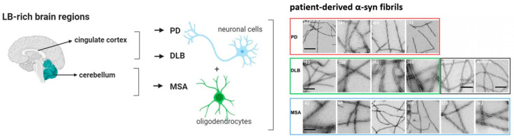
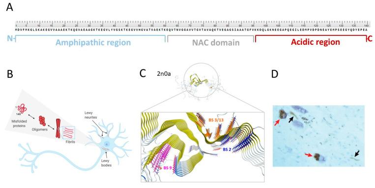
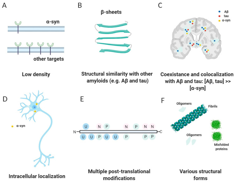
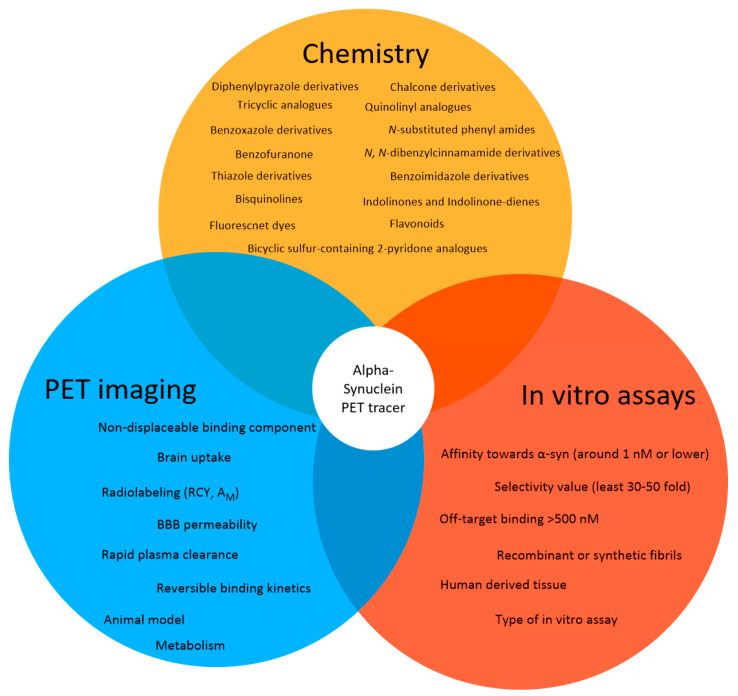
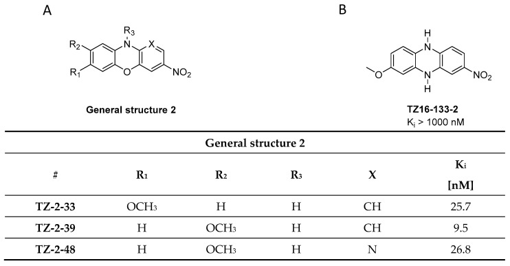
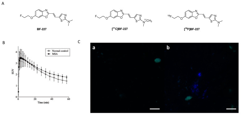
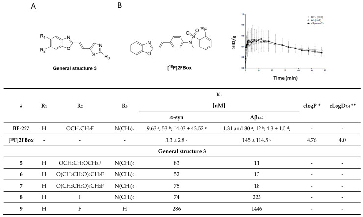
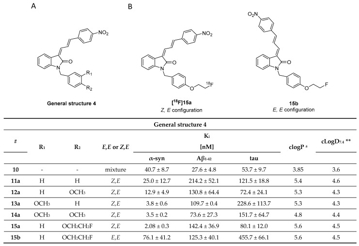
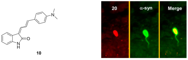
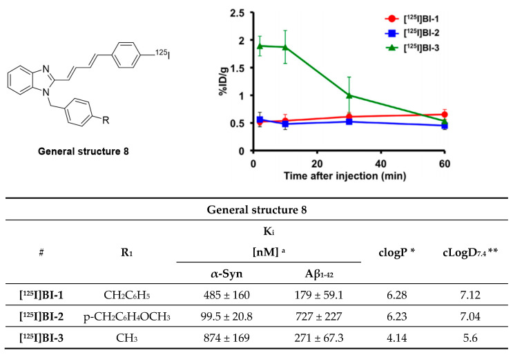
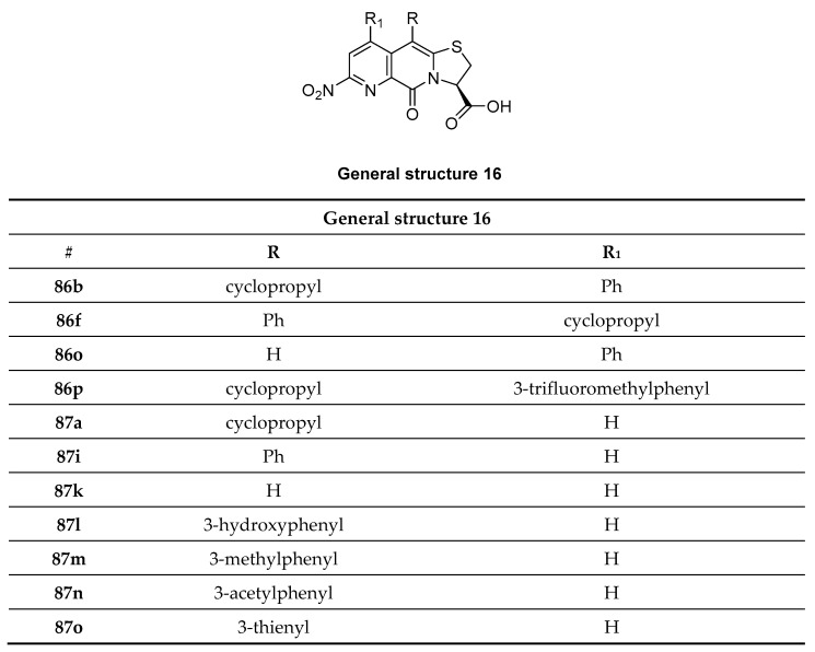
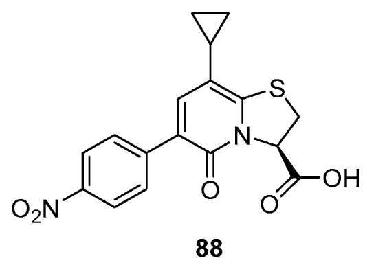
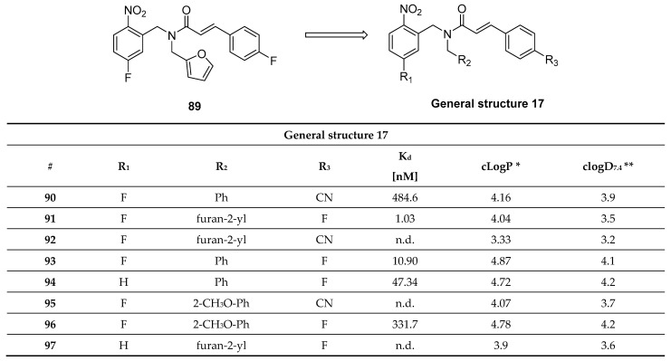
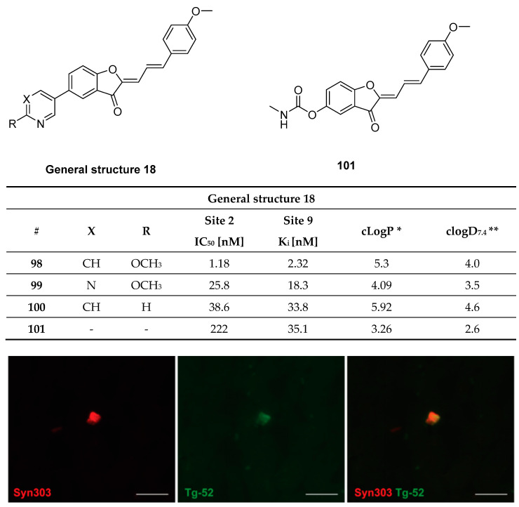
Neurodegenerative diseases such as Parkinson's disease (PD) are manifested by inclusion bodies of alpha-synuclein (α-syn) also called α-synucleinopathies. Detection of these inclusions is thus far only possible by histological examination of postmortem brain tissue. The possibility of non-invasively detecting α-syn will therefore provide valuable insights into the disease progression of α-synucleinopathies. In particular, α-syn imaging can quantify changes in monomeric, oligomeric, and fibrillic α-syn over time and improve early diagnosis of various α-synucleinopathies or monitor treatment progress. Positron emission tomography (PET) is a non-invasive in vivo imaging technique that can quantify target expression and drug occupancies when a suitable tracer exists. As such, novel α-syn PET tracers are highly sought after. The development of an α-syn PET tracer faces several challenges. For example, the low abundance of α-syn within the brain necessitates the development of a high-affinity ligand. Moreover, α-syn depositions are, in contrast to amyloid proteins, predominantly localized intracellularly, limiting their accessibility. Furthermore, another challenge is the ligand selectivity over structurally similar amyloids such as amyloid-beta or tau, which are often co-localized with α-syn pathology. The lack of a defined crystal structure of α-syn has also hindered rational drug and tracer design efforts. Our objective for this review is to provide a comprehensive overview of current efforts in the development of selective α-syn PET tracers.

摘要

帕金森病(PD)等神经退行性疾病表现为α-突触核蛋白(α-syn)包涵体,也称为α-突触核蛋白病。迄今为止,只有通过对死后脑组织进行组织学检查才能检测到这些包涵体。因此,非侵入性检测α-syn的可能性将为α-突触核蛋白病的疾病进展提供有价值的见解。特别是,α-syn成像可以量化单体、寡聚体和纤维状α-syn随时间的变化,并改善各种α-突触核蛋白病的早期诊断或监测治疗进展。正电子发射断层扫描(PET)是一种非侵入性的体内成像技术,当存在合适的示踪剂时,可以量化靶标表达和药物占有率。因此,新型α-syn PET示踪剂备受追捧。α-syn PET示踪剂的开发面临几个挑战。例如,大脑中α-syn的丰度较低,这就需要开发一种高亲和力配体。此外,与淀粉样蛋白不同,α-syn沉积物主要定位于细胞内,限制了它们的可及性。此外,另一个挑战是配体对结构相似的淀粉样蛋白(如β-淀粉样蛋白或tau蛋白)的选择性,这些蛋白通常与α-syn病理共存。α-syn缺乏明确的晶体结构也阻碍了合理的药物和示踪剂设计工作。我们撰写这篇综述的目的是全面概述目前在开发选择性α-syn PET示踪剂方面所做的努力。

https://cdn.ncbi.nlm.nih.gov/pmc/blobs/d22f/8466155/d6c89a06a4c7/pharmaceuticals-14-00847-g033.jpg
https://cdn.ncbi.nlm.nih.gov/pmc/blobs/d22f/8466155/84c2bb57fd99/pharmaceuticals-14-00847-g001.jpg
https://cdn.ncbi.nlm.nih.gov/pmc/blobs/d22f/8466155/38484399e349/pharmaceuticals-14-00847-g002.jpg
https://cdn.ncbi.nlm.nih.gov/pmc/blobs/d22f/8466155/e3515c8d95c4/pharmaceuticals-14-00847-g003.jpg
https://cdn.ncbi.nlm.nih.gov/pmc/blobs/d22f/8466155/b0f232b015b4/pharmaceuticals-14-00847-g004.jpg
https://cdn.ncbi.nlm.nih.gov/pmc/blobs/d22f/8466155/fec55e6e962c/pharmaceuticals-14-00847-g005.jpg
https://cdn.ncbi.nlm.nih.gov/pmc/blobs/d22f/8466155/50cf262d663e/pharmaceuticals-14-00847-g006.jpg
https://cdn.ncbi.nlm.nih.gov/pmc/blobs/d22f/8466155/f1db26a60da6/pharmaceuticals-14-00847-g007.jpg
https://cdn.ncbi.nlm.nih.gov/pmc/blobs/d22f/8466155/f45516ef43f2/pharmaceuticals-14-00847-g008.jpg
https://cdn.ncbi.nlm.nih.gov/pmc/blobs/d22f/8466155/c1cf42b9715e/pharmaceuticals-14-00847-g009.jpg
https://cdn.ncbi.nlm.nih.gov/pmc/blobs/d22f/8466155/7b52f611aefd/pharmaceuticals-14-00847-g010.jpg
https://cdn.ncbi.nlm.nih.gov/pmc/blobs/d22f/8466155/034a5abf7043/pharmaceuticals-14-00847-g011.jpg
https://cdn.ncbi.nlm.nih.gov/pmc/blobs/d22f/8466155/ac7c6324a9a6/pharmaceuticals-14-00847-g012.jpg
https://cdn.ncbi.nlm.nih.gov/pmc/blobs/d22f/8466155/a80aab5d609e/pharmaceuticals-14-00847-g013.jpg
https://cdn.ncbi.nlm.nih.gov/pmc/blobs/d22f/8466155/0ceb4e5083c5/pharmaceuticals-14-00847-g014.jpg
https://cdn.ncbi.nlm.nih.gov/pmc/blobs/d22f/8466155/717f9c52ecaa/pharmaceuticals-14-00847-g015.jpg
https://cdn.ncbi.nlm.nih.gov/pmc/blobs/d22f/8466155/d739355167ff/pharmaceuticals-14-00847-g017.jpg
https://cdn.ncbi.nlm.nih.gov/pmc/blobs/d22f/8466155/401474d2d3f1/pharmaceuticals-14-00847-g018.jpg
https://cdn.ncbi.nlm.nih.gov/pmc/blobs/d22f/8466155/ef15fdf04ff0/pharmaceuticals-14-00847-g019.jpg
https://cdn.ncbi.nlm.nih.gov/pmc/blobs/d22f/8466155/664ad134621b/pharmaceuticals-14-00847-g020.jpg
https://cdn.ncbi.nlm.nih.gov/pmc/blobs/d22f/8466155/29eb489c01f5/pharmaceuticals-14-00847-g021.jpg
https://cdn.ncbi.nlm.nih.gov/pmc/blobs/d22f/8466155/8552f14beb25/pharmaceuticals-14-00847-g022.jpg
https://cdn.ncbi.nlm.nih.gov/pmc/blobs/d22f/8466155/7c47610aad02/pharmaceuticals-14-00847-g023.jpg
https://cdn.ncbi.nlm.nih.gov/pmc/blobs/d22f/8466155/f02c7d2ddabd/pharmaceuticals-14-00847-g024.jpg
https://cdn.ncbi.nlm.nih.gov/pmc/blobs/d22f/8466155/0310fae04de7/pharmaceuticals-14-00847-g025.jpg
https://cdn.ncbi.nlm.nih.gov/pmc/blobs/d22f/8466155/c41cfa84935e/pharmaceuticals-14-00847-g026a.jpg
https://cdn.ncbi.nlm.nih.gov/pmc/blobs/d22f/8466155/3b6bdf074c81/pharmaceuticals-14-00847-g027.jpg
https://cdn.ncbi.nlm.nih.gov/pmc/blobs/d22f/8466155/91fe75472ffa/pharmaceuticals-14-00847-g028.jpg
https://cdn.ncbi.nlm.nih.gov/pmc/blobs/d22f/8466155/a90bd05013a1/pharmaceuticals-14-00847-g029.jpg
https://cdn.ncbi.nlm.nih.gov/pmc/blobs/d22f/8466155/da3568ee4873/pharmaceuticals-14-00847-g030.jpg
https://cdn.ncbi.nlm.nih.gov/pmc/blobs/d22f/8466155/4246637a6166/pharmaceuticals-14-00847-g031.jpg
https://cdn.ncbi.nlm.nih.gov/pmc/blobs/d22f/8466155/198637e5ba61/pharmaceuticals-14-00847-g032.jpg
https://cdn.ncbi.nlm.nih.gov/pmc/blobs/d22f/8466155/d6c89a06a4c7/pharmaceuticals-14-00847-g033.jpg

相似文献

1
Alpha-Synuclein PET Tracer Development-An Overview about Current Efforts.α-突触核蛋白正电子发射断层显像示踪剂的研发——当前研究进展概述
Pharmaceuticals (Basel). 2021 Aug 26;14(9):847. doi: 10.3390/ph14090847.
2
Associations Between APOE Variants, Tau and α-Synuclein.载脂蛋白 E 变异体与 Tau 和 α-突触核蛋白的关联。
Adv Exp Med Biol. 2019;1184:177-186. doi: 10.1007/978-981-32-9358-8_15.
3
Development of an α-synuclein positron emission tomography tracer for imaging synucleinopathies.用于成像突触核蛋白病的α-突触核蛋白正电子发射断层扫描示踪剂的开发。
Cell. 2023 Aug 3;186(16):3350-3367.e19. doi: 10.1016/j.cell.2023.06.004. Epub 2023 Jul 7.
4
Small-molecule PET Tracers for Imaging Proteinopathies.小分子 PET 示踪剂用于成像蛋白病。
Semin Nucl Med. 2017 Sep;47(5):553-575. doi: 10.1053/j.semnuclmed.2017.06.003. Epub 2017 Jul 13.
5
Advancing Parkinson's Disease Diagnostics: The Potential of Arylpyrazolethiazole Derivatives for Imaging α-Synuclein Aggregates.推进帕金森病诊断:芳基吡唑噻唑衍生物用于成像α-突触核蛋白聚集体的潜力
ACS Omega. 2024 May 30;9(23):24774-24788. doi: 10.1021/acsomega.4c01301. eCollection 2024 Jun 11.
6
Toward Novel [F]Fluorine-Labeled Radiotracers for the Imaging of α-Synuclein Fibrils.用于α-突触核蛋白原纤维成像的新型[F]氟标记放射性示踪剂
Front Aging Neurosci. 2022 Apr 29;14:830704. doi: 10.3389/fnagi.2022.830704. eCollection 2022.
7
FKBP12-immunopositive inclusions in patients with α-synucleinopathies.α-突触核蛋白病患者中FKBP12免疫阳性包涵体
Brain Res. 2018 Feb 1;1680:39-45. doi: 10.1016/j.brainres.2017.12.012. Epub 2017 Dec 12.
8
In Silico, in Vitro, and in Vivo Evaluation of New Candidates for α-Synuclein PET Imaging.新型α-突触核蛋白 PET 成像示踪剂的计算机模拟、体外和体内评价。
Mol Pharm. 2018 Aug 6;15(8):3153-3166. doi: 10.1021/acs.molpharmaceut.8b00229. Epub 2018 Jul 25.
9
A simple, versatile and robust centrifugation-based filtration protocol for the isolation and quantification of α-synuclein monomers, oligomers and fibrils: Towards improving experimental reproducibility in α-synuclein research.一种简单、通用且稳健的基于离心的过滤方案,用于分离和定量α-突触核蛋白单体、寡聚体和纤维:提高 α-突触核蛋白研究中的实验可重复性。
J Neurochem. 2020 Apr;153(1):103-119. doi: 10.1111/jnc.14955. Epub 2020 Feb 25.
10
Sensitive fluorescence polarization technique for rapid screening of alpha-synuclein oligomerization/fibrillization inhibitors.用于快速筛选α-突触核蛋白寡聚化/纤维化抑制剂的灵敏荧光偏振技术
Biochemistry. 2007 Nov 6;46(44):12522-9. doi: 10.1021/bi701128c. Epub 2007 Oct 10.

引用本文的文献

1
Current Status of α-Synuclein Biomarkers and the Need for α-Synuclein PET Tracers.α-突触核蛋白生物标志物的现状及对α-突触核蛋白PET示踪剂的需求
Cells. 2025 Aug 18;14(16):1272. doi: 10.3390/cells14161272.
2
The quest for Parkinson's disease biomarkers: traditional and emerging multi-omics approaches.帕金森病生物标志物的探索:传统与新兴的多组学方法
Mol Biol Rep. 2025 Aug 16;52(1):831. doi: 10.1007/s11033-025-10929-x.
3
Resynchronization of microglial activity in the brain is associated with restoration of motor function in Parkinson's disease.

本文引用的文献

1
Identification of a nanomolar affinity α-synuclein fibril imaging probe by ultra-high throughput screening.通过超高通量筛选鉴定纳摩尔亲和力的α-突触核蛋白原纤维成像探针。
Chem Sci. 2020 Sep 10;11(47):12746-12754. doi: 10.1039/d0sc02159h. eCollection 2020 Dec 21.
2
Fluorescent Probe for Selective Imaging of α-Synuclein Fibrils in Living Cells.用于活细胞中 α-突触核蛋白纤维选择性成像的荧光探针。
ACS Chem Neurosci. 2021 Apr 21;12(8):1293-1298. doi: 10.1021/acschemneuro.1c00090. Epub 2021 Apr 5.
3
[C]MODAG-001-towards a PET tracer targeting α-synuclein aggregates.
大脑中微胶质细胞活动的重新同步与帕金森病运动功能的恢复有关。
Commun Biol. 2025 Aug 9;8(1):1188. doi: 10.1038/s42003-025-08602-x.
4
Discovery of -(6-Methoxypyridin-3-yl)quinoline-2-amine Derivatives for Imaging Aggregated α-Synuclein in Parkinson's Disease with Positron Emission Tomography.用于正电子发射断层扫描成像帕金森病中聚集的α-突触核蛋白的-(6-甲氧基吡啶-3-基)喹啉-2-胺衍生物的发现
Cells. 2025 Jul 18;14(14):1108. doi: 10.3390/cells14141108.
5
Detecting alpha-synuclein aggregates with small molecules on single-molecule array.在单分子阵列上用小分子检测α-突触核蛋白聚集体。
Chem Sci. 2025 Jun 16. doi: 10.1039/d4sc07649d.
6
Development of Positron Emission Tomography Radiotracers for Imaging α-Synuclein Aggregates.用于α-突触核蛋白聚集体成像的正电子发射断层显像剂的研发
Cells. 2025 Jun 16;14(12):907. doi: 10.3390/cells14120907.
7
Ligands for Protein Fibrils of Amyloid-β, α-Synuclein, and Tau.淀粉样β蛋白、α-突触核蛋白和tau蛋白原纤维的配体
Chem Rev. 2025 Jun 11;125(11):5282-5348. doi: 10.1021/acs.chemrev.4c00838. Epub 2025 May 6.
8
Alpha-synuclein misfolding as fluid biomarker for Parkinson's disease measured with the iRS platform.使用iRS平台测量α-突触核蛋白错误折叠作为帕金森病的液体生物标志物。
EMBO Mol Med. 2025 Apr 25. doi: 10.1038/s44321-025-00229-z.
9
PET tracer development for imaging α-synucleinopathies.用于成像α-突触核蛋白病的正电子发射断层扫描(PET)示踪剂开发
Arch Pharm Res. 2025 Apr;48(4):333-350. doi: 10.1007/s12272-025-01538-0. Epub 2025 Apr 14.
10
Biomarker-guided decision making in clinical drug development for neurodegenerative disorders.神经退行性疾病临床药物研发中的生物标志物引导决策
Nat Rev Drug Discov. 2025 Apr 4. doi: 10.1038/s41573-025-01165-w.
[C]MODAG-001-一种针对α-突触核蛋白聚集的 PET 示踪剂。
Eur J Nucl Med Mol Imaging. 2021 Jun;48(6):1759-1772. doi: 10.1007/s00259-020-05133-x. Epub 2020 Dec 28.
4
Identification and Evaluation of Bisquinoline Scaffold as a New Candidate for α-Synuclein-PET Imaging.鉴定和评估双喹啉支架作为一种新的α-突触核蛋白-PET 成像候选物。
ACS Chem Neurosci. 2020 Dec 16;11(24):4254-4261. doi: 10.1021/acschemneuro.0c00523. Epub 2020 Dec 1.
5
Fluorine-18-Labeled Antibody Ligands for PET Imaging of Amyloid-β in Brain.氟-18 标记的抗体配体用于脑内淀粉样蛋白-β的 PET 成像。
ACS Chem Neurosci. 2020 Dec 16;11(24):4460-4468. doi: 10.1021/acschemneuro.0c00652. Epub 2020 Nov 25.
6
On the consensus nomenclature rules for radiopharmaceutical chemistry - Reconsideration of radiochemical conversion.放射性药物化学共识命名规则 - 放射性化学转化的再思考。
Nucl Med Biol. 2021 Feb;93:19-21. doi: 10.1016/j.nucmedbio.2020.11.003. Epub 2020 Nov 18.
7
Identification and in vitro characterization of C05-01, a PBB3 derivative with improved affinity for alpha-synuclein.鉴定并体外研究 C05-01,一种与α-突触核蛋白亲和力提高的 PBB3 衍生物。
Brain Res. 2020 Dec 15;1749:147131. doi: 10.1016/j.brainres.2020.147131. Epub 2020 Sep 18.
8
Novel Furan-2-yl-1-pyrazoles Possess Inhibitory Activity against α-Synuclein Aggregation.新型呋喃-2-基-1-吡唑对α-突触核蛋白聚集具有抑制活性。
ACS Chem Neurosci. 2020 Aug 5;11(15):2303-2315. doi: 10.1021/acschemneuro.0c00252. Epub 2020 Jul 8.
9
Structures of α-synuclein filaments from multiple system atrophy.多系统萎缩中α-突触核蛋白丝的结构。
Nature. 2020 Sep;585(7825):464-469. doi: 10.1038/s41586-020-2317-6. Epub 2020 May 27.
10
The structural differences between patient-derived α-synuclein strains dictate characteristics of Parkinson's disease, multiple system atrophy and dementia with Lewy bodies.患者源性 α-突触核蛋白菌株之间的结构差异决定了帕金森病、多系统萎缩和路易体痴呆的特征。
Acta Neuropathol. 2020 Jun;139(6):977-1000. doi: 10.1007/s00401-020-02157-3. Epub 2020 Apr 30.