Department of Orthodontics, Shanghai Ninth People's Hospital, College of Stomatology, Shanghai Jiao Tong University School of Medical, Shanghai, 200011, China.
BMC Oral Health. 2021 Sep 27;21(1):475. doi: 10.1186/s12903-021-01839-y.
The aim of the study was to analyze the morphology and position of the tongue and hyoid bone in skeletal Class II patients with different vertical growth patterns by cone beam computed tomography in comparison to skeletal Class I patients.
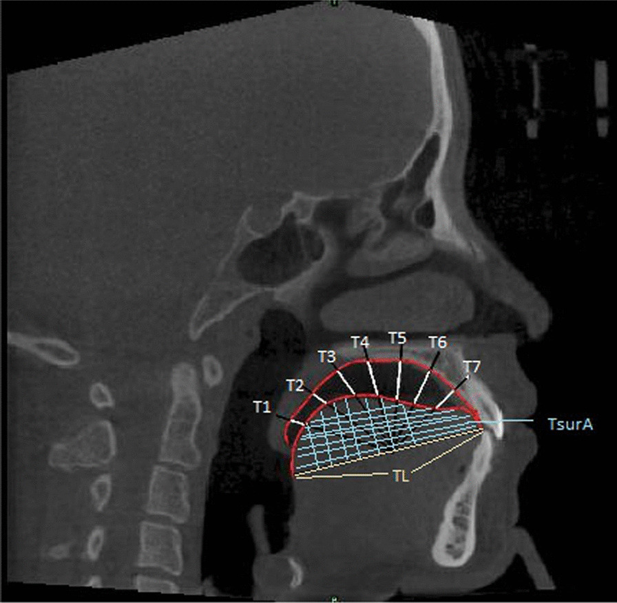
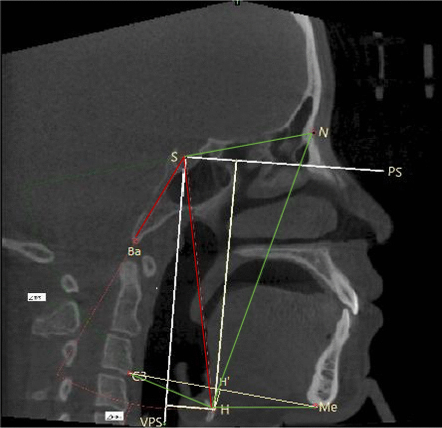
Ninety subjects with malocclusion were divided into skeletal Class II and Class I groups by ANB angles. Based on different vertical growth patterns, subjects in each group were divided into 3 subgroups: high-angle group (MP-FH ≥ 32.0°), average-angle group (22.0° ≤ MP-FH < 32°) and low-angle group (MP-FH < 22°). The position and morphology of the tongue and hyoid bone were evaluated in the cone beam computed tomography images. The independent Student's t-test was used to compare the position and morphology of the tongue and hyoid bone between skeletal Class I and Class II groups. One-way analysis of variance (ANOVA) was used to compare the measurement indexes of different vertical facial patterns in each group.
Patients in skeletal Class II group had lower tongue posture, and the tongue body was smaller than that of those in the Class I group (P < 0.05). The position of the hyoid bone was lower in the skeletal Class II group than in Class I group (P < 0.05). The tongue length and H-Me in the skeletal Class I group with a low angle were significantly larger than those with an average angle and high angle (P < 0.05). There was no significant difference in the position or morphology of the tongue and hyoid bone in the skeletal Class II group with different vertical facial patterns (P > 0.05).
Patients with skeletal Class II malocclusion have lower tongue posture, a smaller tongue body, and greater occurrence of posterior inferior hyoid bone position than skeletal Class I patients. The length of the mandibular body in skeletal Class I patients with a horizontal growth type is longer. The position and morphology of the tongue and hyoid bone were not greatly affected by vertical facial development in skeletal Class II patients.
本研究旨在通过锥形束 CT 分析不同垂直生长型骨性Ⅱ类患者与骨性Ⅰ类患者的舌体及舌骨形态和位置。
90 例错颌畸形患者按 ANB 角分为骨性Ⅱ类组和骨性Ⅰ类组。根据不同的垂直生长型,每组患者再分为 3 个亚组:高角组(MP-FH≥32.0°)、均角组(22.0°≤MP-FH<32°)和低角组(MP-FH<22°)。在锥形束 CT 图像上评估舌体及舌骨的位置和形态。采用独立样本 t 检验比较骨性Ⅰ类组和骨性Ⅱ类组舌体及舌骨的位置和形态。采用单因素方差分析比较各组不同垂直面型的测量指标。
骨性Ⅱ类组患者的舌位较低,舌体较小(P<0.05)。骨性Ⅱ类组患者的舌骨位置较骨性Ⅰ类组低(P<0.05)。骨性Ⅰ类组低角患者的舌长和 H-Me 显著大于均角组和高角组(P<0.05)。骨性Ⅱ类组不同垂直面型患者的舌体及舌骨位置或形态无显著差异(P>0.05)。
骨性Ⅱ类错颌患者的舌位较低,舌体较小,舌后下骨位置较骨性Ⅰ类患者更常见。骨性Ⅰ类水平生长型患者的下颌体长。骨性Ⅱ类患者的垂直面型发育对舌体及舌骨的位置和形态影响不大。