• 文献检索
  • 文档翻译
  • 深度研究
  • 学术资讯
  • Suppr Zotero 插件Zotero 插件
  • 邀请有礼
  • 套餐&价格
  • 历史记录
应用&插件
Suppr Zotero 插件Zotero 插件浏览器插件Mac 客户端Windows 客户端微信小程序
定价
高级版会员购买积分包购买API积分包
服务
文献检索文档翻译深度研究API 文档MCP 服务
关于我们
关于 Suppr公司介绍联系我们用户协议隐私条款
关注我们

Suppr 超能文献

核心技术专利:CN118964589B侵权必究
粤ICP备2023148730 号-1Suppr @ 2026

文献检索

告别复杂PubMed语法,用中文像聊天一样搜索,搜遍4000万医学文献。AI智能推荐,让科研检索更轻松。

立即免费搜索

文件翻译

保留排版,准确专业,支持PDF/Word/PPT等文件格式,支持 12+语言互译。

免费翻译文档

深度研究

AI帮你快速写综述,25分钟生成高质量综述,智能提取关键信息,辅助科研写作。

立即免费体验

表面形貌指数:一种用于漏斗胸畸形严重程度评估的新指标。

Surface topography index: a novel deformity severity assessment index for pectus excavatum.

作者信息

Wang Hui, Liu Wei, Zhang Dong-Ying, Si Wen-Yue, Yang Qing-Lin, Lu Lian-Wei, Wang Feng-Hua, Li Le, Wang Qi, Xia Hui-Min

机构信息

The Second School of Clinical Medicine, Southern Medical University, Guangzhou, China.

Department of Pediatric Surgery, Guangzhou Women and Children's Medical Center, Guangzhou Medical University, Guangzhou, China.

出版信息

Transl Pediatr. 2021 Aug;10(8):2044-2051. doi: 10.21037/tp-21-282.

DOI:10.21037/tp-21-282
PMID:34584874
原文链接:https://pmc.ncbi.nlm.nih.gov/articles/PMC8429865/
Abstract

BACKGROUND

The surface topography index (STI) has great potential in both routine computed tomography (CT) scan and emerging optical imaging systems. However, the diagnostic accuracy and stability of the STI as a deformity severity assessment index has not been fully confirmed. Therefore, the aim of the present study was to determine the diagnostic performance of the STI as a novel deformity severity assessment index for pectus excavatum.

METHODS

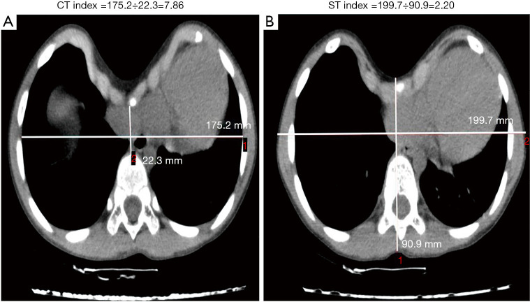
The present study consisted of 722 chest CT images from a single center. The standard CT index (CTI) and STI were calculated for all patients. The between-group difference and the level of compliance between the CTI and STI was analyzed by -test and Pearson correlation. The diagnostic value and optimum discriminatory values of the CTI and STI were calculated by a receiver-operating characteristic (ROC) curve and DeLong's test.

RESULTS

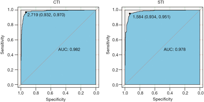
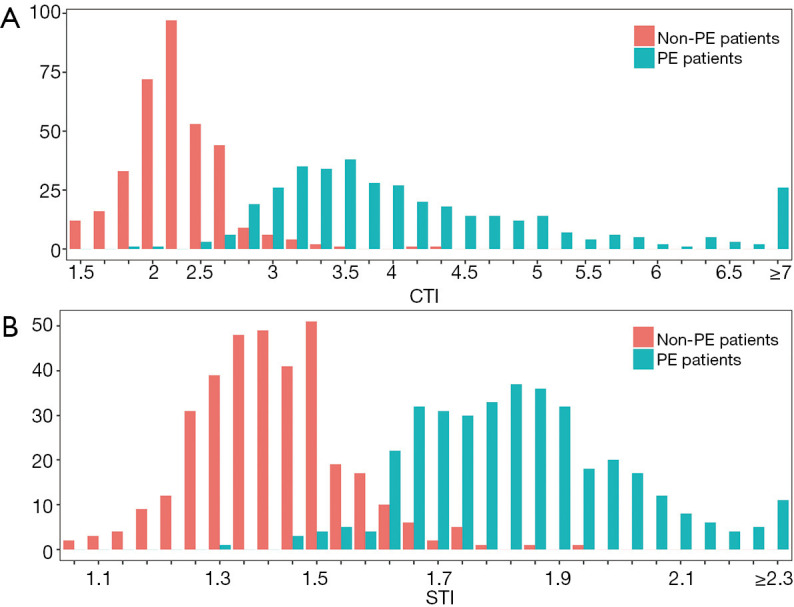
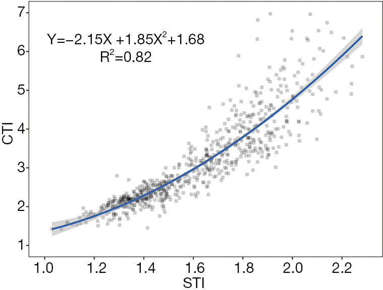
The distributions of the CTI and STI were similar and showed a slight overlap between the pectus excavatum (PE) and non-PE groups. Both the CTI and STI significantly differed between the 2 groups (P<0.001). The STI demonstrated a strong Pearson correlation with the CTI (r=0.91, 95% confidence interval: 0.88-0.91, P<0.001). The ROC curves showed that STI =1.58 (sensitivity: 0.93, specificity: 0.95) could be considered equivalent to CTI =2.72 (sensitivity: 0.93, specificity: 0.97) as the optimum discriminatory values. DeLong's test showed no significant difference in the ROC curve results between the CTI and STI (Z=0.90, P=0.37).

CONCLUSIONS

The STI has comparative discrimination ability in PE diagnosis and deformity severity assessment when used with the standard CTI. The STI as a novel index is not only an ideal evaluation metric of PE deformity but also an objective trait for PE patients just as weight and height for everyone.

摘要

背景

表面形貌指数(STI)在常规计算机断层扫描(CT)和新兴光学成像系统中均具有巨大潜力。然而,作为畸形严重程度评估指标,STI的诊断准确性和稳定性尚未得到充分证实。因此,本研究旨在确定STI作为漏斗胸畸形严重程度评估新指标的诊断性能。

方法

本研究纳入来自单一中心的722例胸部CT图像。计算所有患者的标准CT指数(CTI)和STI。通过t检验和Pearson相关性分析CTI与STI之间的组间差异和一致性水平。采用受试者操作特征(ROC)曲线和DeLong检验计算CTI和STI的诊断价值及最佳鉴别值。

结果

CTI和STI的分布相似,漏斗胸(PE)组与非PE组之间存在轻微重叠。两组间CTI和STI均有显著差异(P<0.001)。STI与CTI呈强Pearson相关性(r=0.91,95%置信区间:0.88-0.91,P<0.001)。ROC曲线显示,STI =1.58(敏感性:0.93,特异性:0.95)可被视为等同于CTI =2.72(敏感性:0.93,特异性:0.97)的最佳鉴别值。DeLong检验显示CTI和STI的ROC曲线结果无显著差异(Z=0.90,P=0.37)。

结论

与标准CTI联合使用时,STI在PE诊断和畸形严重程度评估方面具有相当的鉴别能力。作为一个新指标,STI不仅是PE畸形的理想评估指标,也是PE患者的一个客观特征,就如同体重和身高对每个人一样。

https://cdn.ncbi.nlm.nih.gov/pmc/blobs/28b8/8429865/b985f8a9b0fc/tp-10-08-2044-f4.jpg
https://cdn.ncbi.nlm.nih.gov/pmc/blobs/28b8/8429865/daec3b7363cf/tp-10-08-2044-f1.jpg
https://cdn.ncbi.nlm.nih.gov/pmc/blobs/28b8/8429865/e321504ddf80/tp-10-08-2044-f2.jpg
https://cdn.ncbi.nlm.nih.gov/pmc/blobs/28b8/8429865/db72dd7d4cf1/tp-10-08-2044-f3.jpg
https://cdn.ncbi.nlm.nih.gov/pmc/blobs/28b8/8429865/b985f8a9b0fc/tp-10-08-2044-f4.jpg

相似文献

1
Surface topography index: a novel deformity severity assessment index for pectus excavatum.表面形貌指数:一种用于漏斗胸畸形严重程度评估的新指标。
Transl Pediatr. 2021 Aug;10(8):2044-2051. doi: 10.21037/tp-21-282.
2
Comparison of Haller index values calculated with chest radiographs versus CT for pectus excavatum evaluation.比较采用胸部 X 光片和 CT 计算的 Haller 指数值在漏斗胸评估中的应用。
Pediatr Radiol. 2010 Nov;40(11):1763-7. doi: 10.1007/s00247-010-1681-z. Epub 2010 May 15.
3
Evaluation of the Nuss procedure using pre- and postoperative computed tomographic index.使用术前和术后计算机断层扫描指数评估努斯手术。
J Pediatr Surg. 2008 Mar;43(3):518-21. doi: 10.1016/j.jpedsurg.2007.10.033.
4
The modified percent depth: Another step toward quantifying severity of pectus excavatum without cross-sectional imaging.改良深度百分比:迈向无需横断面成像即可量化漏斗胸严重程度的又一步。
J Pediatr Surg. 2017 Jul;52(7):1098-1101. doi: 10.1016/j.jpedsurg.2017.01.053. Epub 2017 Jan 31.
5
External caliper-based measurements of the modified percent depth as an alternative to cross-sectional imaging for assessing the severity of pectus excavatum.基于外部卡尺测量改良深度百分比,作为评估漏斗胸严重程度的横断面成像替代方法。
J Pediatr Surg. 2020 Jun;55(6):1058-1064. doi: 10.1016/j.jpedsurg.2020.02.053. Epub 2020 Feb 27.
6
Measuring the impact of surgical intervention on pediatric pectus excavatum using white light scanning.使用白光扫描测量手术干预对小儿漏斗胸的影响。
J Pediatr Surg. 2019 Nov;54(11):2261-2267. doi: 10.1016/j.jpedsurg.2019.01.007. Epub 2019 Jan 23.
7
Development of new cardiac deformity indexes for pectus excavatum on computed tomography: feasibility for pre- and post-operative evaluation.计算机断层扫描中漏斗胸新心脏畸形指数的开发:术前和术后评估的可行性
Yonsei Med J. 2009 Jun 30;50(3):385-90. doi: 10.3349/ymj.2009.50.3.385. Epub 2009 Jun 23.
8
A novel technique to measure severity of pediatric pectus excavatum using white light scanning.一种使用白光扫描测量小儿漏斗胸严重程度的新技术。
J Pediatr Surg. 2019 Apr;54(4):656-662. doi: 10.1016/j.jpedsurg.2018.04.017. Epub 2018 Apr 18.
9
Clinical impact of optical imaging with 3-D reconstruction of torso topography in common anterior chest wall anomalies.三维重建胸部体表形态的光学成像在常见前胸壁畸形中的临床应用
J Pediatr Surg. 2007 May;42(5):898-903. doi: 10.1016/j.jpedsurg.2006.12.070.
10
Physician-Estimated Depth as a Screening Tool for Computed Tomography Evaluation of Pectus Excavatum.医生估计深度作为漏斗胸计算机断层扫描评估的筛选工具。
J Surg Res. 2020 Dec;256:687-692. doi: 10.1016/j.jss.2020.03.072. Epub 2020 May 22.

引用本文的文献

1
Advancements in preoperative imaging of pectus excavatum: a comprehensive review.漏斗胸术前影像学进展:全面综述
J Thorac Dis. 2024 Jan 30;16(1):696-707. doi: 10.21037/jtd-23-662. Epub 2024 Jan 24.

本文引用的文献

1
Chest X-ray Dose Equivalent Low-dose CT with Tin Filtration: Potential Role for the Assessment of Pectus Excavatum.胸部 X 射线剂量当量 低剂量 CT 与锡滤过:评估漏斗胸的潜在作用。
Acad Radiol. 2020 May;27(5):644-650. doi: 10.1016/j.acra.2019.07.017. Epub 2019 Aug 28.
2
Surgical management of pectus excavatum in China: results of a survey amongst members of the Chinese Association of Thoracic Surgeons.中国漏斗胸的外科治疗:中国胸外科医师协会成员的调查结果
Ann Transl Med. 2019 May;7(9):202. doi: 10.21037/atm.2019.05.03.
3
Three-Dimensional Optical Imaging for Pectus Excavatum Assessment.
三维光学成像在漏斗胸评估中的应用。
Ann Thorac Surg. 2019 Oct;108(4):1065-1071. doi: 10.1016/j.athoracsur.2019.04.074. Epub 2019 Jun 12.
4
A novel technique to measure severity of pediatric pectus excavatum using white light scanning.一种使用白光扫描测量小儿漏斗胸严重程度的新技术。
J Pediatr Surg. 2019 Apr;54(4):656-662. doi: 10.1016/j.jpedsurg.2018.04.017. Epub 2018 Apr 18.
5
Acceptability, Precision and Accuracy of 3D Photonic Scanning for Measurement of Body Shape in a Multi-Ethnic Sample of Children Aged 5-11 Years: The SLIC Study.5至11岁多民族儿童样本中用于身体形状测量的3D光子扫描的可接受性、精确性和准确性:SLIC研究
PLoS One. 2015 Apr 28;10(4):e0124193. doi: 10.1371/journal.pone.0124193. eCollection 2015.
6
Leonardo da Vinci and the first portrayal of pectus excavatum.
Thorax. 2013 Nov;68(11):1081. doi: 10.1136/thoraxjnl-2013-203224. Epub 2013 Feb 6.
7
Cardiac magnetic resonance imaging for perioperative evaluation of sternal eversion for pectus excavatum.心脏磁共振成像在漏斗胸胸骨翻转术围手术期评估中的应用。
Eur J Cardiothorac Surg. 2013 Jun;43(6):1110-3. doi: 10.1093/ejcts/ezs662. Epub 2013 Jan 7.
8
Comparison of Haller index values calculated with chest radiographs versus CT for pectus excavatum evaluation.比较采用胸部 X 光片和 CT 计算的 Haller 指数值在漏斗胸评估中的应用。
Pediatr Radiol. 2010 Nov;40(11):1763-7. doi: 10.1007/s00247-010-1681-z. Epub 2010 May 15.
9
Method of pectus excavatum measurement based on structured light technique.
J Biomed Opt. 2009 Jul-Aug;14(4):044041. doi: 10.1117/1.3210782.
10
Pectus excavatum: historical background, clinical picture, preoperative evaluation and criteria for operation.漏斗胸:历史背景、临床表现、术前评估及手术标准
Semin Pediatr Surg. 2008 Aug;17(3):181-93. doi: 10.1053/j.sempedsurg.2008.03.002.