Department of Gastroenterology and Hepatology, Osaka University Graduate School of Medicine, Osaka, Japan.
Department of Medicine, Columbia University, New York, NY, USA.
Hepatol Commun. 2022 Feb;6(2):411-422. doi: 10.1002/hep4.1815. Epub 2021 Sep 28.
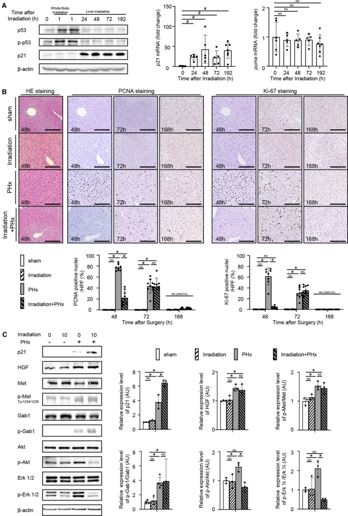
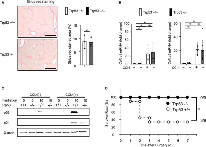
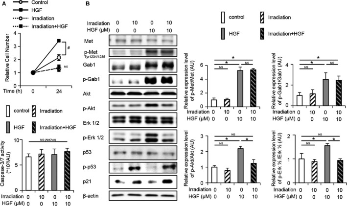
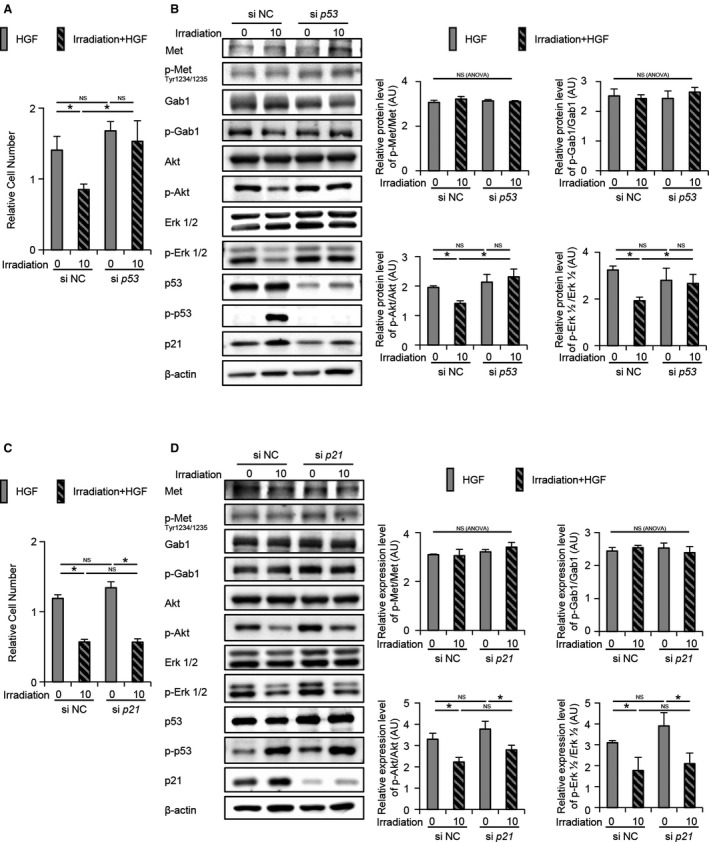
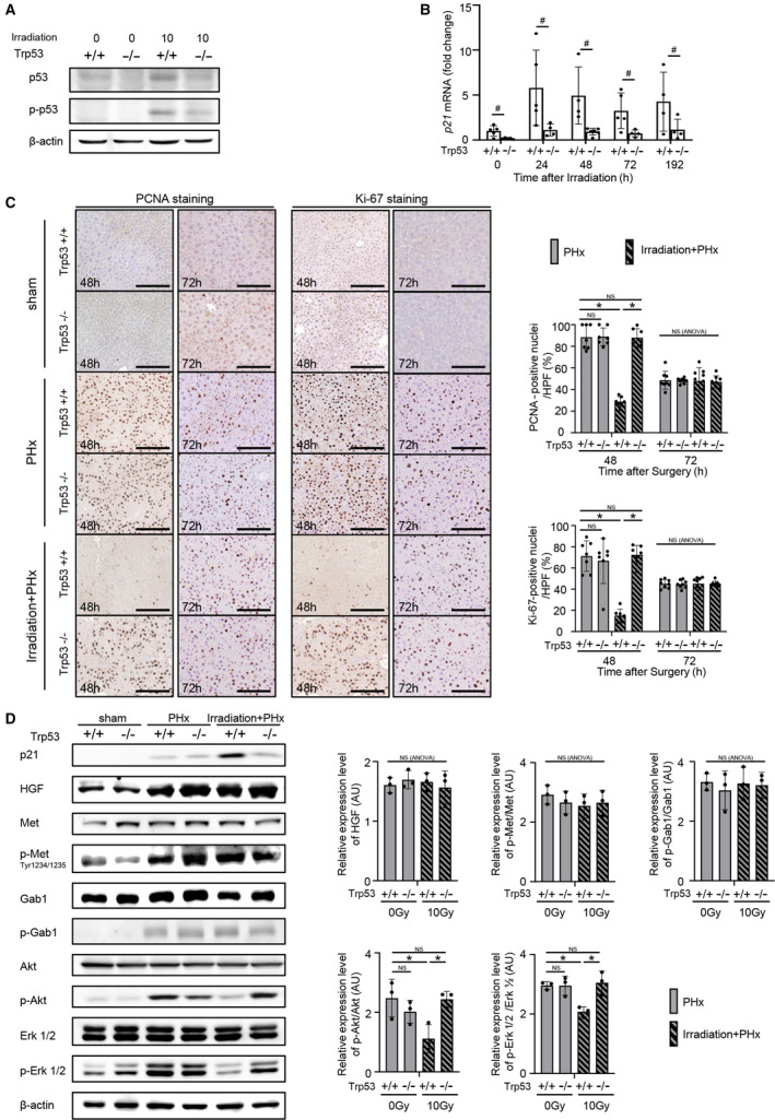
Radiation therapy is one of the treatment methods for hepatocellular carcinoma. However, radiation tolerance of the liver is low, and the detailed effect of radiation on liver regeneration has not been clarified. C57BL/6J mice or hepatocyte-specific p53 knockout (KO) mice (albumin [Alb]-Cre Trp53 ) were irradiated with a single fraction of 10 Gy localized to the upper abdomen. We performed 70% partial hepatectomy (PHx) 24 hours after irradiation. Liver regeneration was assessed by proliferation cell nuclear antigen (PCNA)- and Ki-67-positive hepatocyte ratios and liver-to-body weight ratio after PHx. To establish a fibrosis model, CCl4 was orally administered for 8 weeks. The murine hepatocyte cell line BNL CL.2 (CL2) was irradiated with 10 Gy. Irradiation activated p53, induced downstream p21 in the liver, and delayed liver regeneration after PHx. While PHx increased hepatocyte growth factor (HGF) levels and activated Met with or without irradiation in the regenerative liver, it activated Akt and extracellular kinase 1 and 2 (Erk 1/2) less in irradiated mice than in nonirradiated mice. In CL2 cells cultured with HGF, irradiation suppressed cell growth by decreasing phosphorylated Akt and Erk 1/2 levels, which was abolished by small interfering RNA-mediated p53 knockdown but not by p21 knockdown. Hepatocyte-specific knockout of p53 in mice abolished the irradiation-induced suppression of both liver regeneration and Akt and Erk 1/2 activation after PHx. In the fibrotic mouse model, the survival rate after PHx of irradiated p53 KO mice was higher than that of wild-type mice. Conclusion: p53 but not p21 is involved in the impaired regenerative ability of the irradiated liver.
放射治疗是肝癌的治疗方法之一。然而,肝脏对辐射的耐受性较低,并且辐射对肝再生的详细影响尚未阐明。用 10Gy 的单次剂量局部照射 C57BL/6J 小鼠或肝细胞特异性 p53 敲除(KO)小鼠(白蛋白 [Alb]-Cre Trp53 )。照射后 24 小时进行 70%部分肝切除术(PHx)。通过增殖细胞核抗原(PCNA)-和 Ki-67 阳性肝细胞比例以及 PHx 后的肝体比评估肝再生。为了建立纤维化模型,用 CCl4 口服给药 8 周。用 10Gy 照射小鼠肝细胞系 BNL CL.2(CL2)。照射激活了 p53,在 PHx 后诱导了肝脏中的下游 p21,并延迟了肝再生。虽然 PHx 在再生肝脏中增加了肝细胞生长因子(HGF)水平并激活了 Met,无论是否照射,但在照射小鼠中,其激活 Akt 和细胞外激酶 1 和 2(Erk 1/2)的程度低于非照射小鼠。在 CL2 细胞中用 HGF 培养时,照射通过降低磷酸化 Akt 和 Erk 1/2 水平来抑制细胞生长,这种抑制作用可被小干扰 RNA 介导的 p53 敲低消除,但不能被 p21 敲低消除。在小鼠中敲除肝细胞特异性 p53 可消除照射后 PHx 对肝再生和 Akt 和 Erk 1/2 激活的抑制作用。在纤维化小鼠模型中,照射的 p53 KO 小鼠在 PHx 后的存活率高于野生型小鼠。结论:p53 而不是 p21 参与了照射肝脏再生能力受损。