Section of Digestive Diseases, Department of Internal Medicine, Yale University School of Medicine, New Haven, CT, USA.
Hepatology. 2013 May;57(5):1992-2003. doi: 10.1002/hep.26235. Epub 2013 Mar 19.
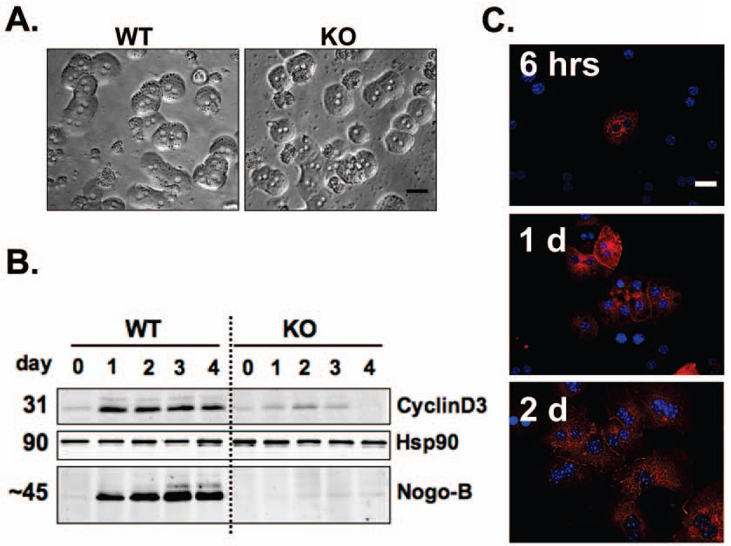
Nogo-B, also known as reticulon 4B, promotes liver fibrosis and cirrhosis by facilitating the transforming growth factor β (TGF-β) signaling pathway in activated hepatic stellate cells. The aim of this study was to determine the role of Nogo-B in hepatocyte proliferation and liver regeneration. Partial hepatectomy (PHx, 70% resection) was performed in male wild-type (WT) and Nogo-A/B knockout mice (referred to as Nogo-B KO mice). Remnant livers were isolated 2 hours, 5 hours, and 1, 2, 3, 7, and 14 days after PHx. Hepatocyte proliferation was assessed by Ki67 labeling index. Quantitative real-time polymerase chain reaction was performed for genes known to be involved in liver regeneration. Hepatocytes isolated from WT and Nogo-B KO mice were used to examine the role of Nogo-B in interleukin-6 (IL-6), hepatocyte growth factor (HGF), epidermal growth factor (EGF), and TGF-β signaling. Nogo-B protein levels increased in the regenerating livers in a time-dependent manner after PHx. Specifically, Nogo-B expression in hepatocytes gradually spread from the periportal toward the central areas by 7 days after PHx, but receded notably by 14 days. Nogo-B facilitated IL-6/signal transducer and activator of transcription 3 signaling, increased HGF-induced but not EGF-induced hepatocyte proliferation, and tended to reduce TGF-β1-induced suppression of hepatocyte proliferation in cultured hepatocytes. Lack of Nogo-B significantly induced TGF-β1 and inhibitor of DNA binding expression 1 day after PHx and IL-6 and EGF expression 2 days after PHx. Lack of Nogo-B delayed hepatocyte proliferation but did not affect the liver-to-body ratio in the regenerative process.
Nogo-B expression in hepatocytes facilitates hepatocyte proliferation and liver regeneration.
Nogo-B,也称为 reticulon 4B,通过促进活化的肝星状细胞中的转化生长因子 β(TGF-β)信号通路,促进肝纤维化和肝硬化。本研究旨在确定 Nogo-B 在肝细胞增殖和肝再生中的作用。对雄性野生型(WT)和 Nogo-A/B 敲除小鼠(称为 Nogo-B KO 小鼠)进行部分肝切除术(PHx,70%切除)。在 PHx 后 2 小时、5 小时以及 1、2、3、7 和 14 天分离残余肝脏。通过 Ki67 标记指数评估肝细胞增殖。对已知参与肝再生的基因进行实时定量聚合酶链反应。使用从 WT 和 Nogo-B KO 小鼠分离的肝细胞来研究 Nogo-B 在白细胞介素 6(IL-6)、肝细胞生长因子(HGF)、表皮生长因子(EGF)和 TGF-β信号中的作用。PHx 后,Nogo-B 蛋白水平在再生肝脏中呈时间依赖性增加。具体而言,Nogo-B 在 PHx 后 7 天内逐渐从门脉周围向中央区域扩散,但在 14 天内明显消退。Nogo-B 促进了 IL-6/信号转导和转录激活因子 3 信号,增加了 HGF 诱导但不增加 EGF 诱导的肝细胞增殖,并倾向于减少 TGF-β1 诱导的肝细胞增殖抑制作用在培养的肝细胞中。缺乏 Nogo-B 显著诱导 PHx 后 1 天 TGF-β1 和 DNA 结合抑制因子 1 的表达,以及 PHx 后 2 天 IL-6 和 EGF 的表达。缺乏 Nogo-B 延迟了肝细胞增殖,但不影响再生过程中的肝体比。
肝细胞中 Nogo-B 的表达促进了肝细胞增殖和肝再生。