Ambrose Natalie, Rodriguez Michael, Waters Karen A, Machaalani Rita
Discipline of Medicine, Central Clinical School, Faculty of Medicine and Health, The University of Sydney, NSW, 2006, Australia.
Department of Pathology, School of Medical Sciences, Faculty of Medicine and Health, The University of Sydney, NSW, 2006, Australia.
Brain Behav Immun Health. 2020 Jul 25;7:100117. doi: 10.1016/j.bbih.2020.100117. eCollection 2020 Aug.
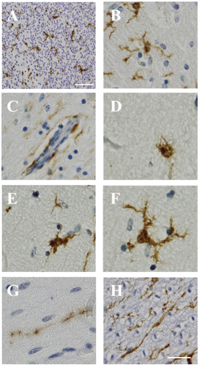
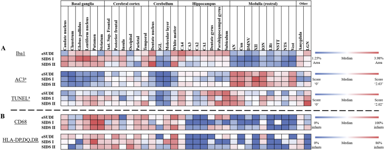
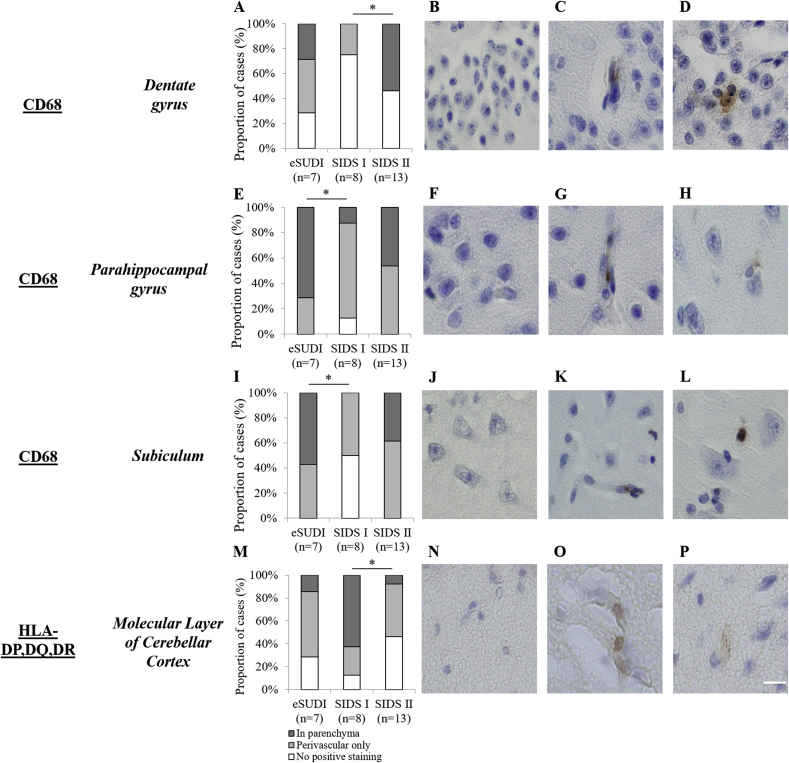
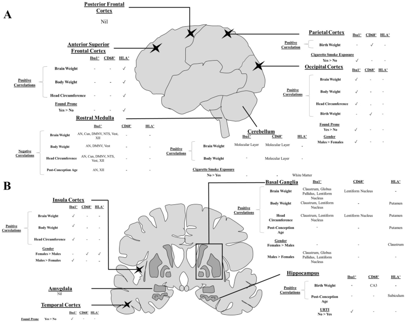
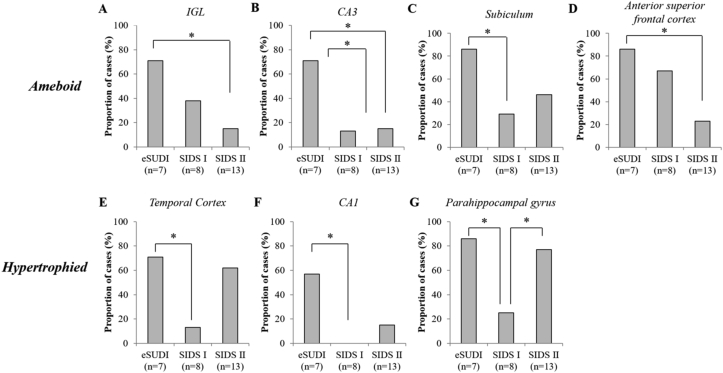
The present study reports on the microglial populations present in 34 regions of the human infant brain (1-11 months), and whether developmental parameters or extrinsic factors such as cigarette smoke exposure, prone sleeping and an upper respiratory tract infection (URTI) influence their expression. Further, we compare microglia populations amongst three sudden unexpected death in infancy (SUDI) sub-groups: explained SUDI (eSUDI, n = 7), sudden infant death syndrome (SIDS) I (n = 8) and SIDS II (n = 13). Ionised calcium binding adaptor molecule-1 (Iba1) was used to determine the morphology and area covered by microglia in a given brain region. Activation was explored using cluster-of-differentiation factor 68 (CD68) and human leukocyte antigen-DP,DQ,DR (HLA). We found regional heterogeneity in the area covered and activation status of microglia across the infant brain. The hippocampus, basal ganglia, white matter and dentate nucleus of the cerebellum showed larger areas of Iba1, while the brainstem had the smallest. Microglia in regions of the basal ganglia and cortex demonstrated positive correlations with infant developmental parameters, while in nuclei of the rostral medulla, negative correlations between microglia parameters were seen. URTI and cigarette smoke exposure were associated with a reduced microglial area in regions of the hippocampus and cortex (parietal and occipital), respectively. In the context of SIDS, a reduced microglial area was seen in SIDS II and fewer SIDS I infants demonstrated activated phenotypes in the hippocampus. Overall, we identify the distribution of microglia in the infant brain to be heterogenous, and influenced by intrinsic and extrinsic factors, and that the SIDS I group is a useful control group for future research into other infant CNS pathologies.
本研究报告了人类婴儿大脑34个区域(1 - 11个月)中存在的小胶质细胞群体,以及发育参数或外部因素,如接触香烟烟雾、俯卧睡眠和上呼吸道感染(URTI)是否会影响它们的表达。此外,我们比较了婴儿猝死综合征(SUDI)三个亚组中的小胶质细胞群体:可解释的SUDI(eSUDI,n = 7)、婴儿猝死综合征(SIDS)I(n = 8)和SIDS II(n = 13)。使用离子钙结合衔接分子-1(Iba1)来确定给定脑区中小胶质细胞覆盖的形态和面积。使用分化簇因子68(CD68)和人类白细胞抗原-DP、DQ、DR(HLA)来探究激活情况。我们发现婴儿大脑中小胶质细胞覆盖面积和激活状态存在区域异质性。海马体、基底神经节、白质和小脑齿状核显示出较大的Iba1面积,而脑干的面积最小。基底神经节和皮质区域的小胶质细胞与婴儿发育参数呈正相关,而在延髓前部核中,小胶质细胞参数之间呈负相关。URTI和接触香烟烟雾分别与海马体和皮质(顶叶和枕叶)区域中小胶质细胞面积减少有关。在SIDS的背景下,SIDS II中小胶质细胞面积减少,且较少有SIDS I婴儿在海马体中表现出激活表型。总体而言,我们确定婴儿大脑中小胶质细胞的分布是异质性的,并且受内在和外在因素影响,而且SIDS I组是未来研究其他婴儿中枢神经系统疾病的有用对照组。