• 文献检索
  • 文档翻译
  • 深度研究
  • 学术资讯
  • Suppr Zotero 插件Zotero 插件
  • 邀请有礼
  • 套餐&价格
  • 历史记录
应用&插件
Suppr Zotero 插件Zotero 插件浏览器插件Mac 客户端Windows 客户端微信小程序
定价
高级版会员购买积分包购买API积分包
服务
文献检索文档翻译深度研究API 文档MCP 服务
关于我们
关于 Suppr公司介绍联系我们用户协议隐私条款
关注我们

Suppr 超能文献

核心技术专利:CN118964589B侵权必究
粤ICP备2023148730 号-1Suppr @ 2026

文献检索

告别复杂PubMed语法,用中文像聊天一样搜索,搜遍4000万医学文献。AI智能推荐,让科研检索更轻松。

立即免费搜索

文件翻译

保留排版,准确专业,支持PDF/Word/PPT等文件格式,支持 12+语言互译。

免费翻译文档

深度研究

AI帮你快速写综述,25分钟生成高质量综述,智能提取关键信息,辅助科研写作。

立即免费体验

小鼠初级运动皮层的细胞解剖结构。

Cellular anatomy of the mouse primary motor cortex.

机构信息

Cold Spring Harbor Laboratory, Cold Spring Harbor, NY, USA.

UCLA Brain Research and Artificial Intelligence Nexus, Department of Neurobiology, David Geffen School of Medicine, University of California Los Angeles, Los Angeles, CA, USA.

出版信息

Nature. 2021 Oct;598(7879):159-166. doi: 10.1038/s41586-021-03970-w. Epub 2021 Oct 6.

DOI:10.1038/s41586-021-03970-w
PMID:34616071
原文链接:https://pmc.ncbi.nlm.nih.gov/articles/PMC8494646/
Abstract

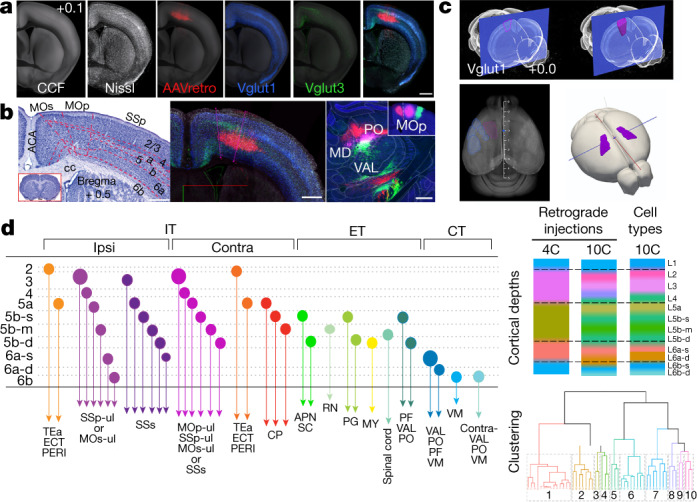
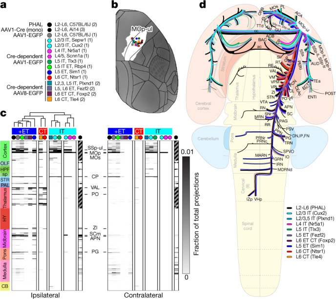
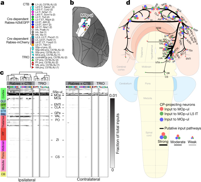
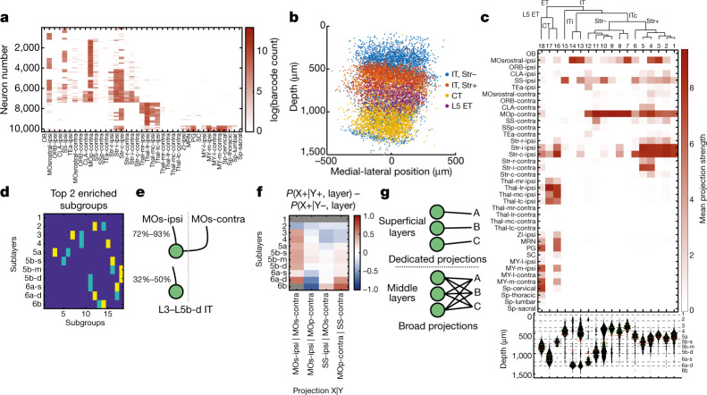
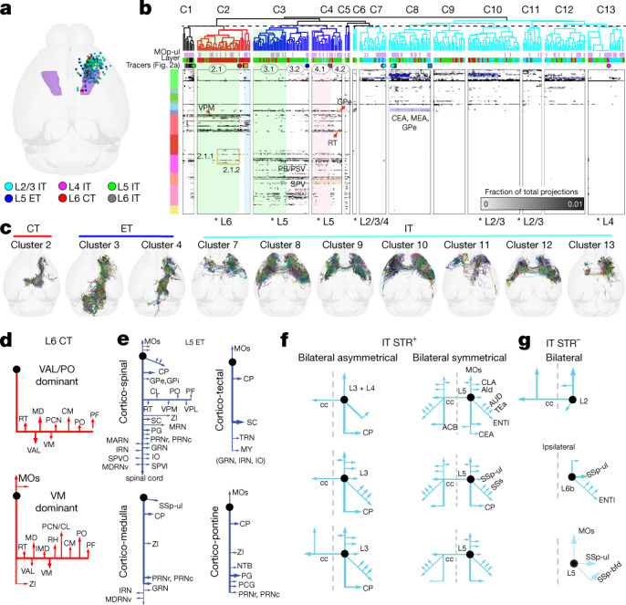
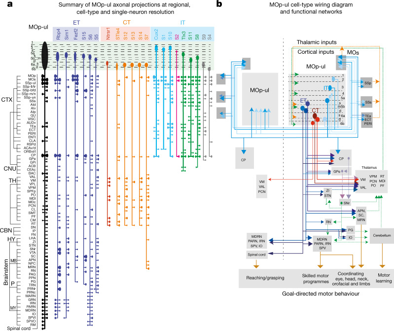
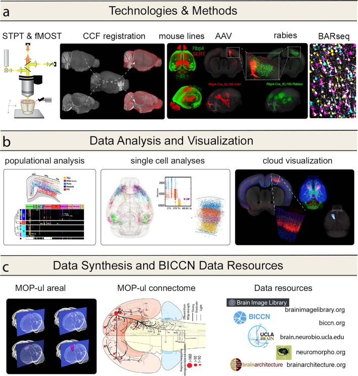
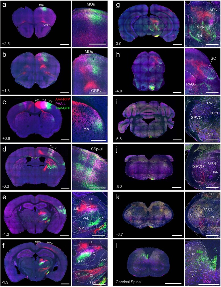
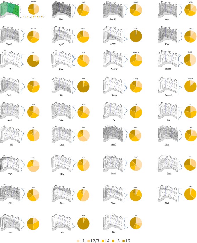
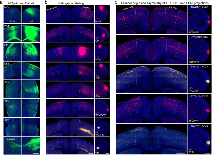
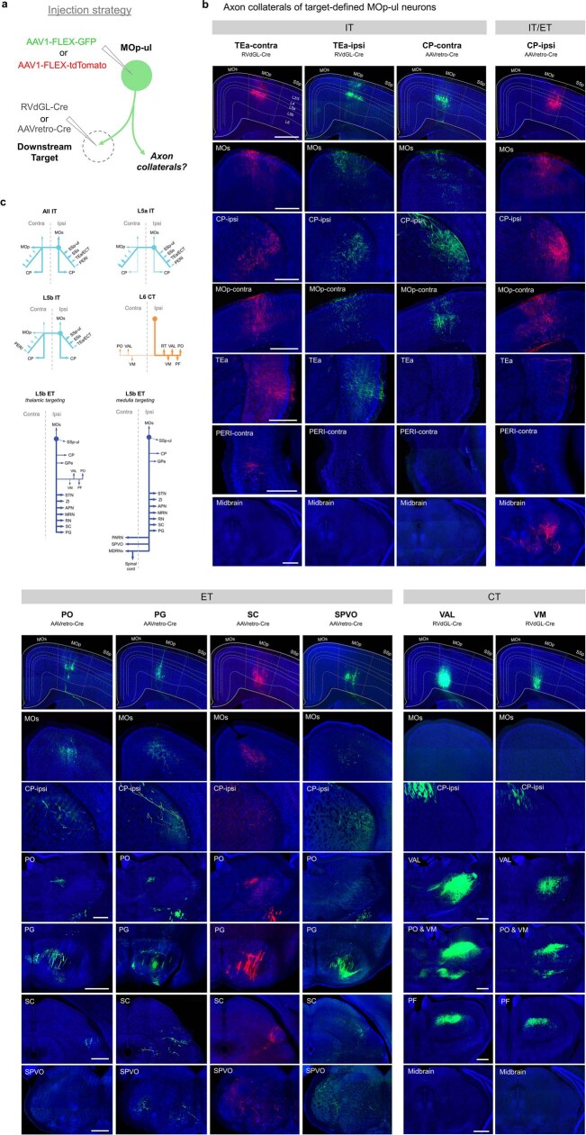
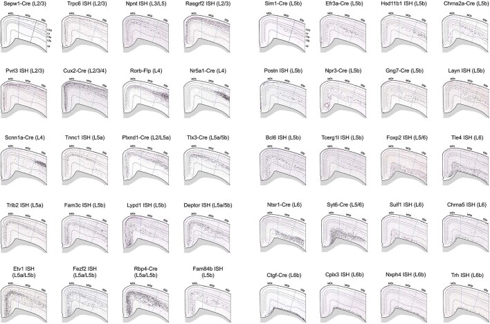
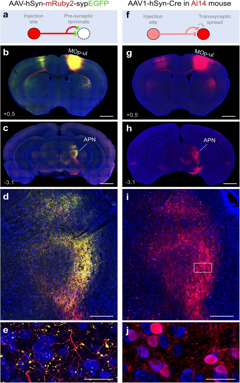
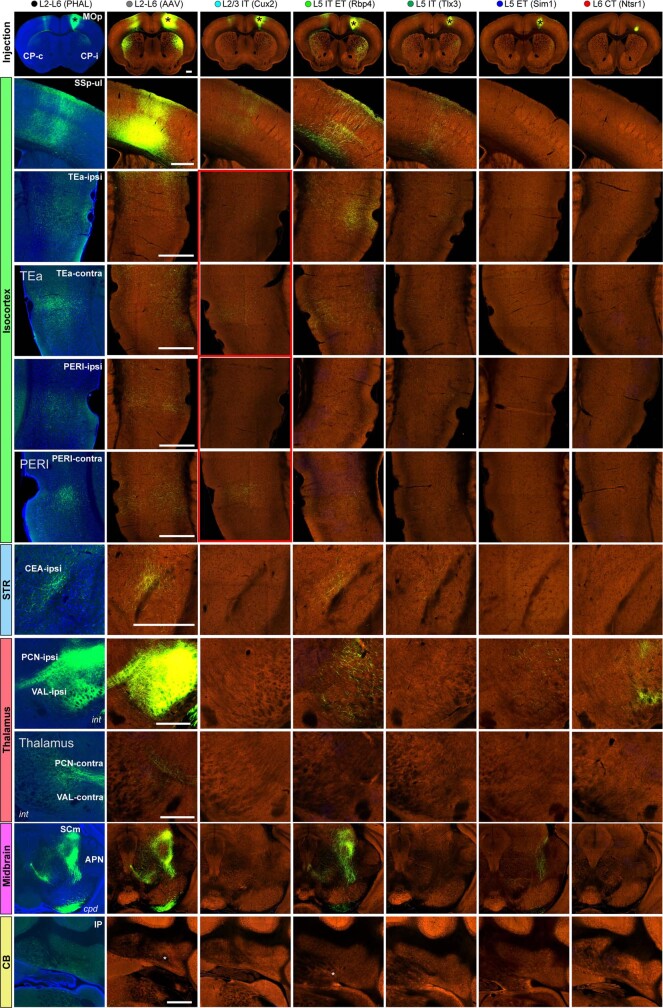
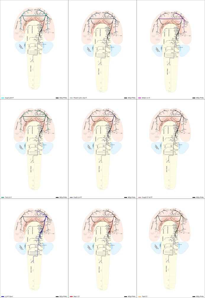
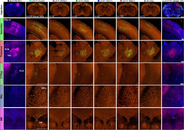
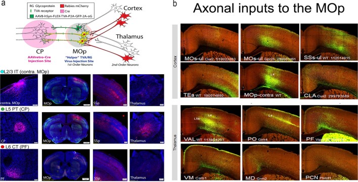
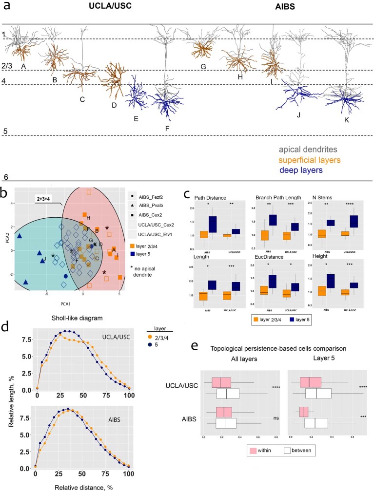
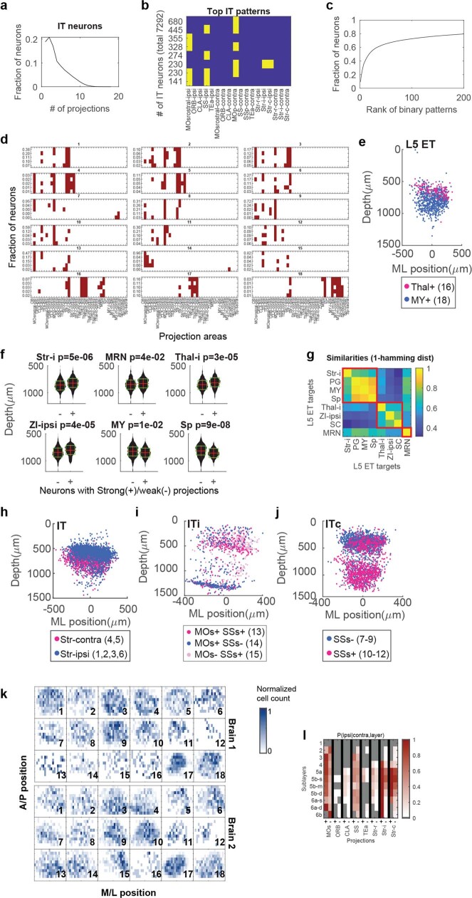
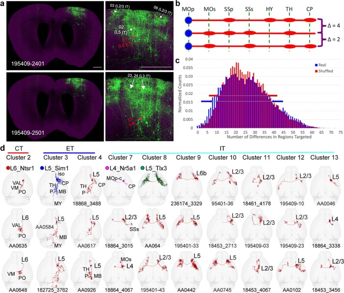
An essential step toward understanding brain function is to establish a structural framework with cellular resolution on which multi-scale datasets spanning molecules, cells, circuits and systems can be integrated and interpreted. Here, as part of the collaborative Brain Initiative Cell Census Network (BICCN), we derive a comprehensive cell type-based anatomical description of one exemplar brain structure, the mouse primary motor cortex, upper limb area (MOp-ul). Using genetic and viral labelling, barcoded anatomy resolved by sequencing, single-neuron reconstruction, whole-brain imaging and cloud-based neuroinformatics tools, we delineated the MOp-ul in 3D and refined its sublaminar organization. We defined around two dozen projection neuron types in the MOp-ul and derived an input-output wiring diagram, which will facilitate future analyses of motor control circuitry across molecular, cellular and system levels. This work provides a roadmap towards a comprehensive cellular-resolution description of mammalian brain architecture.

摘要

理解大脑功能的重要步骤是建立一个具有细胞分辨率的结构框架,在此基础上可以整合和解释跨越分子、细胞、回路和系统的多尺度数据集。在这里,作为合作的大脑倡议细胞普查网络 (BICCN) 的一部分,我们对一个范例脑结构,即小鼠初级运动皮层上肢区 (MOp-ul),进行了基于细胞类型的全面解剖描述。使用遗传和病毒标记、通过测序解析的条形码解剖、单细胞重建、全脑成像和基于云的神经信息学工具,我们对 MOp-ul 进行了 3D 描绘,并细化了其亚层组织。我们在 MOp-ul 中定义了大约二十几种投射神经元类型,并得出了一个输入-输出接线图,这将有助于未来在分子、细胞和系统水平上对运动控制回路进行分析。这项工作为哺乳动物大脑结构的全面细胞分辨率描述提供了路线图。

https://cdn.ncbi.nlm.nih.gov/pmc/blobs/54ef/8494646/1a9a25f514cc/41586_2021_3970_Fig27_ESM.jpg
https://cdn.ncbi.nlm.nih.gov/pmc/blobs/54ef/8494646/46f04bc22f90/41586_2021_3970_Fig1_HTML.jpg
https://cdn.ncbi.nlm.nih.gov/pmc/blobs/54ef/8494646/63ed7d899146/41586_2021_3970_Fig2_HTML.jpg
https://cdn.ncbi.nlm.nih.gov/pmc/blobs/54ef/8494646/5a012c70e725/41586_2021_3970_Fig3_HTML.jpg
https://cdn.ncbi.nlm.nih.gov/pmc/blobs/54ef/8494646/6fa7faac8c40/41586_2021_3970_Fig4_HTML.jpg
https://cdn.ncbi.nlm.nih.gov/pmc/blobs/54ef/8494646/f866e9aadafd/41586_2021_3970_Fig5_HTML.jpg
https://cdn.ncbi.nlm.nih.gov/pmc/blobs/54ef/8494646/acde2bfdb777/41586_2021_3970_Fig6_HTML.jpg
https://cdn.ncbi.nlm.nih.gov/pmc/blobs/54ef/8494646/a1525e7ca7c4/41586_2021_3970_Fig7_ESM.jpg
https://cdn.ncbi.nlm.nih.gov/pmc/blobs/54ef/8494646/068e705709b4/41586_2021_3970_Fig8_ESM.jpg
https://cdn.ncbi.nlm.nih.gov/pmc/blobs/54ef/8494646/f2d9ea0e5afe/41586_2021_3970_Fig9_ESM.jpg
https://cdn.ncbi.nlm.nih.gov/pmc/blobs/54ef/8494646/c98097aa769b/41586_2021_3970_Fig10_ESM.jpg
https://cdn.ncbi.nlm.nih.gov/pmc/blobs/54ef/8494646/ba27322cc398/41586_2021_3970_Fig11_ESM.jpg
https://cdn.ncbi.nlm.nih.gov/pmc/blobs/54ef/8494646/c420f08edbab/41586_2021_3970_Fig12_ESM.jpg
https://cdn.ncbi.nlm.nih.gov/pmc/blobs/54ef/8494646/a1af6af2b4f1/41586_2021_3970_Fig13_ESM.jpg
https://cdn.ncbi.nlm.nih.gov/pmc/blobs/54ef/8494646/9a5f03ce5767/41586_2021_3970_Fig14_ESM.jpg
https://cdn.ncbi.nlm.nih.gov/pmc/blobs/54ef/8494646/78c429f570e5/41586_2021_3970_Fig15_ESM.jpg
https://cdn.ncbi.nlm.nih.gov/pmc/blobs/54ef/8494646/c36df75937ea/41586_2021_3970_Fig16_ESM.jpg
https://cdn.ncbi.nlm.nih.gov/pmc/blobs/54ef/8494646/485cfefe9d34/41586_2021_3970_Fig17_ESM.jpg
https://cdn.ncbi.nlm.nih.gov/pmc/blobs/54ef/8494646/277f28da63d4/41586_2021_3970_Fig18_ESM.jpg
https://cdn.ncbi.nlm.nih.gov/pmc/blobs/54ef/8494646/c0f3f2fe26d4/41586_2021_3970_Fig19_ESM.jpg
https://cdn.ncbi.nlm.nih.gov/pmc/blobs/54ef/8494646/2ef3946905e3/41586_2021_3970_Fig20_ESM.jpg
https://cdn.ncbi.nlm.nih.gov/pmc/blobs/54ef/8494646/faca0de182b7/41586_2021_3970_Fig21_ESM.jpg
https://cdn.ncbi.nlm.nih.gov/pmc/blobs/54ef/8494646/19a47a336d10/41586_2021_3970_Fig22_ESM.jpg
https://cdn.ncbi.nlm.nih.gov/pmc/blobs/54ef/8494646/fabde1f5c193/41586_2021_3970_Fig23_ESM.jpg
https://cdn.ncbi.nlm.nih.gov/pmc/blobs/54ef/8494646/9f3ac5258561/41586_2021_3970_Fig24_ESM.jpg
https://cdn.ncbi.nlm.nih.gov/pmc/blobs/54ef/8494646/a5ecd35d9780/41586_2021_3970_Fig25_ESM.jpg
https://cdn.ncbi.nlm.nih.gov/pmc/blobs/54ef/8494646/e64b4c750919/41586_2021_3970_Fig26_ESM.jpg
https://cdn.ncbi.nlm.nih.gov/pmc/blobs/54ef/8494646/1a9a25f514cc/41586_2021_3970_Fig27_ESM.jpg

相似文献

1
Cellular anatomy of the mouse primary motor cortex.小鼠初级运动皮层的细胞解剖结构。
Nature. 2021 Oct;598(7879):159-166. doi: 10.1038/s41586-021-03970-w. Epub 2021 Oct 6.
2
Spatially resolved cell atlas of the mouse primary motor cortex by MERFISH.通过 MERFISH 技术对小鼠初级运动皮层进行空间分辨的细胞图谱分析。
Nature. 2021 Oct;598(7879):137-143. doi: 10.1038/s41586-021-03705-x. Epub 2021 Oct 6.
3
A multimodal cell census and atlas of the mammalian primary motor cortex.哺乳动物初级运动皮层的多模态细胞普查和图谱
Nature. 2021 Oct;598(7879):86-102. doi: 10.1038/s41586-021-03950-0. Epub 2021 Oct 6.
4
Isoform cell-type specificity in the mouse primary motor cortex.小鼠初级运动皮层中的同工型细胞类型特异性。
Nature. 2021 Oct;598(7879):195-199. doi: 10.1038/s41586-021-03969-3. Epub 2021 Oct 6.
5
Comparative cellular analysis of motor cortex in human, marmoset and mouse.人类、狨猴和小鼠运动皮层的比较细胞分析。
Nature. 2021 Oct;598(7879):111-119. doi: 10.1038/s41586-021-03465-8. Epub 2021 Oct 6.
6
Phenotypic variation of transcriptomic cell types in mouse motor cortex.小鼠运动皮层转录组细胞类型的表型变异。
Nature. 2021 Oct;598(7879):144-150. doi: 10.1038/s41586-020-2907-3. Epub 2020 Nov 12.
7
A transcriptomic and epigenomic cell atlas of the mouse primary motor cortex.小鼠初级运动皮层的转录组和表观基因组细胞图谱
Nature. 2021 Oct;598(7879):103-110. doi: 10.1038/s41586-021-03500-8. Epub 2021 Oct 6.
8
Shared and distinct transcriptomic cell types across neocortical areas.不同脑区共有的和独特的转录组细胞类型。
Nature. 2018 Nov;563(7729):72-78. doi: 10.1038/s41586-018-0654-5. Epub 2018 Oct 31.
9
DNA methylation atlas of the mouse brain at single-cell resolution.单细胞分辨率下的小鼠大脑 DNA 甲基化图谱。
Nature. 2021 Oct;598(7879):120-128. doi: 10.1038/s41586-020-03182-8. Epub 2021 Oct 6.
10
Morphological diversity of single neurons in molecularly defined cell types.分子定义的细胞类型中单神经元的形态多样性。
Nature. 2021 Oct;598(7879):174-181. doi: 10.1038/s41586-021-03941-1. Epub 2021 Oct 6.

引用本文的文献

1
Automatic and accurate reconstruction of long-range axonal projections of single-neuron in mouse brain.小鼠大脑中单个神经元长程轴突投射的自动精确重建
Elife. 2025 Sep 1;13:RP102840. doi: 10.7554/eLife.102840.
2
A whole-brain male mouse atlas of long-range inputs to histaminergic neurons.一份关于组胺能神经元长程输入的全脑雄性小鼠图谱。
Nat Commun. 2025 Aug 29;16(1):8092. doi: 10.1038/s41467-025-63394-2.
3
Neural networks of the mouse visceromotor cortex.小鼠内脏运动皮层的神经网络。

本文引用的文献

1
A multimodal cell census and atlas of the mammalian primary motor cortex.哺乳动物初级运动皮层的多模态细胞普查和图谱
Nature. 2021 Oct;598(7879):86-102. doi: 10.1038/s41586-021-03950-0. Epub 2021 Oct 6.
2
Morphological diversity of single neurons in molecularly defined cell types.分子定义的细胞类型中单神经元的形态多样性。
Nature. 2021 Oct;598(7879):174-181. doi: 10.1038/s41586-021-03941-1. Epub 2021 Oct 6.
3
Genetic dissection of the glutamatergic neuron system in cerebral cortex.皮层谷氨酸能神经元系统的遗传剖析。
Nature. 2025 Aug 27. doi: 10.1038/s41586-025-09360-w.
4
A neuronal imaging dataset for deep learning in the reconstruction of single-neuron axons.用于单神经元轴突重建深度学习的神经元成像数据集。
Front Neuroinform. 2025 Aug 5;19:1628030. doi: 10.3389/fninf.2025.1628030. eCollection 2025.
5
Stochastic activity in low-rank recurrent neural networks.低秩递归神经网络中的随机活动。
PLoS Comput Biol. 2025 Aug 18;21(8):e1013371. doi: 10.1371/journal.pcbi.1013371.
6
Motor learning drives region-specific transcriptomic remodeling in the motor cortex and dorsal striatum.运动学习驱动运动皮层和背侧纹状体中特定区域的转录组重塑。
bioRxiv. 2025 Jul 17:2025.07.11.664268. doi: 10.1101/2025.07.11.664268.
7
Distinct neurogenic pathways shape the diversification and mosaic organization of cortical output channels.不同的神经源性通路塑造了皮质输出通道的多样化和镶嵌式组织。
bioRxiv. 2025 Jul 23:2025.07.18.665624. doi: 10.1101/2025.07.18.665624.
8
Long-range cortical GABA neurons oppose binge drinking.远程皮质γ-氨基丁酸能神经元抑制暴饮行为。
Nat Neurosci. 2025 Jul 23. doi: 10.1038/s41593-025-02030-0.
9
Stochastic activity in low-rank recurrent neural networks.低秩递归神经网络中的随机活动。
bioRxiv. 2025 Jul 11:2025.04.22.649933. doi: 10.1101/2025.04.22.649933.
10
Integrated Classification of Cortical Cells and Quantitative Projectomic Mapping Unveil Organizational Principles of Brain-Wide Connectomes at Single Cell Level.皮质细胞的综合分类与定量投射组学图谱揭示单细胞水平全脑连接组的组织原则。
bioRxiv. 2025 May 8:2025.05.08.652699. doi: 10.1101/2025.05.08.652699.
Nature. 2021 Oct;598(7879):182-187. doi: 10.1038/s41586-021-03955-9. Epub 2021 Oct 6.
4
Spatially resolved cell atlas of the mouse primary motor cortex by MERFISH.通过 MERFISH 技术对小鼠初级运动皮层进行空间分辨的细胞图谱分析。
Nature. 2021 Oct;598(7879):137-143. doi: 10.1038/s41586-021-03705-x. Epub 2021 Oct 6.
5
Integrating barcoded neuroanatomy with spatial transcriptional profiling enables identification of gene correlates of projections.将条形码神经解剖学与空间转录组学相结合,可实现对投射相关基因的鉴定。
Nat Neurosci. 2021 Jun;24(6):873-885. doi: 10.1038/s41593-021-00842-4. Epub 2021 May 10.
6
Visualizing anatomically registered data with brainrender.使用 brainrender 可视化解剖配准数据。
Elife. 2021 Mar 19;10:e65751. doi: 10.7554/eLife.65751.
7
Operations Research Methods for Estimating the Population Size of Neuron Types.用于估计神经元类型种群大小的运筹学方法
Ann Oper Res. 2020 Jun;289(1):33-50. doi: 10.1007/s10479-020-03542-7. Epub 2020 Mar 9.
8
Linking axon morphology to gene expression: a strategy for neuronal cell-type classification.将轴突形态与基因表达联系起来:神经元细胞类型分类的策略。
Curr Opin Neurobiol. 2020 Dec;65:70-76. doi: 10.1016/j.conb.2020.10.006. Epub 2020 Nov 9.
9
Brainwide Genetic Sparse Cell Labeling to Illuminate the Morphology of Neurons and Glia with Cre-Dependent MORF Mice.全脑遗传稀疏细胞标记揭示 Cre 依赖型 MORF 小鼠神经元和神经胶质细胞的形态。
Neuron. 2020 Oct 14;108(1):111-127.e6. doi: 10.1016/j.neuron.2020.07.019. Epub 2020 Aug 13.
10
An open access mouse brain flatmap and upgraded rat and human brain flatmaps based on current reference atlases.基于当前参考图谱的开放获取鼠标大脑平铺图和升级的大鼠和人类大脑平铺图。
J Comp Neurol. 2021 Feb;529(3):576-594. doi: 10.1002/cne.24966. Epub 2020 Jul 19.