Department of Nutrition and Integrative Physiology, Molecular Medicine Program, University of Utah, Salt Lake City, UT, USA.
Departments of Biology, Bioengineering, and Medicinal Chemistry, University of Utah, Salt Lake City, UT, USA.
Methods Mol Biol. 2022;2303:495-511. doi: 10.1007/978-1-0716-1398-6_40.
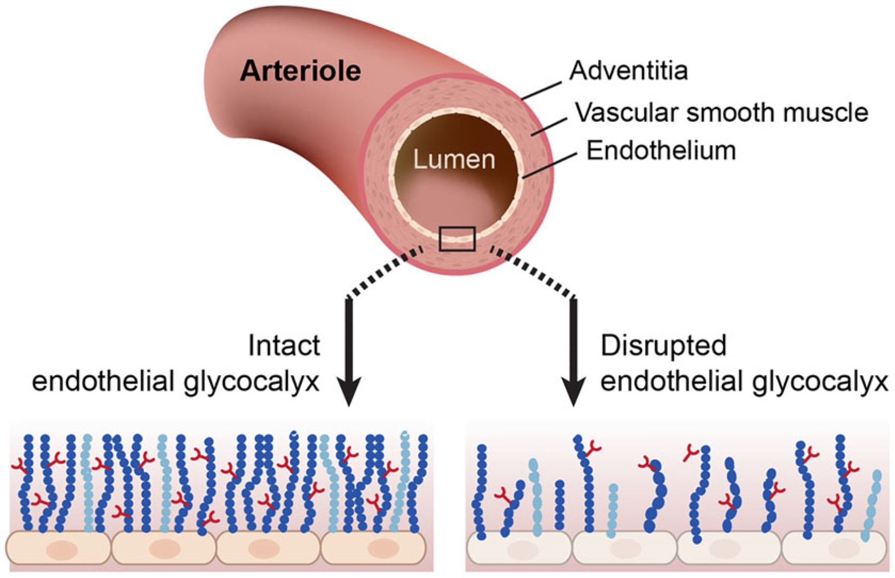
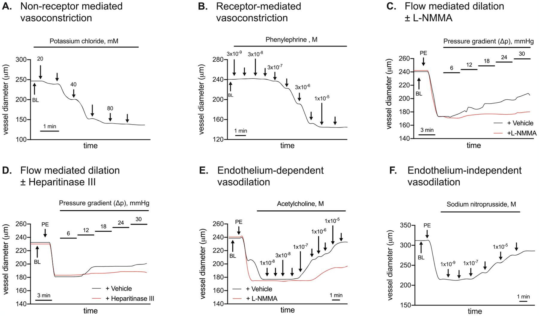
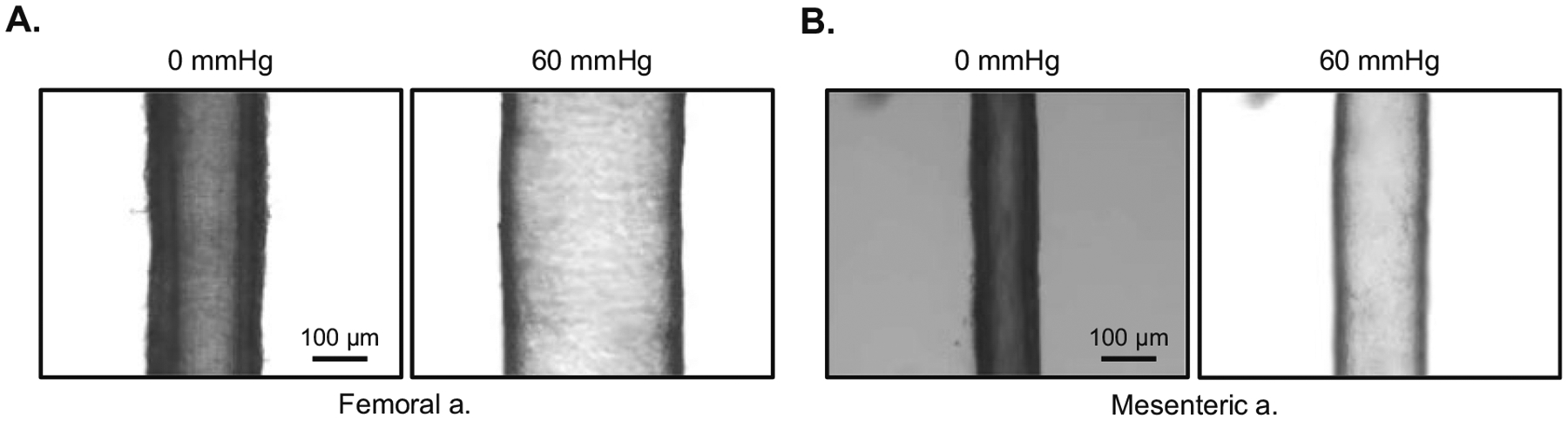
Evidence is emerging that disruption of the endothelial glycocalyx might contribute importantly to arterial dysfunction in the context of diabetes. One approach to assess the integrity of the endothelium and the vascular smooth muscle cell layer, in the absence of neural, humoral, and mechanical influences, is by measuring arterial vasomotion ex vivo. Here we describe a procedure to assess non-receptor-mediated vasoconstriction, receptor-mediated vasoconstriction, and endothelium-dependent and -independent vasodilation, in resistance and conductance arteries pressurized to 60 mmHg. In addition to evaluating vasoreactivity using isobaric approaches, the same experimental set-up can be used to initiate a pressure gradient across the artery such that intraluminal, flow-mediated vasodilation can be measured. After recording endothelium-dependent vasodilation using isobaric or flow-mediated approaches, identical interventions can be completed in the presence of enzymes that cleave biologically active heparan sulfates into inactive disaccharide and oligosaccharide fragments to assess the contribution from: (a) endothelial-derived substances (e.g., nitric oxide via nitric oxide synthase inhibition); or (b) important components of the glycocalyx (e.g., removal of heparan sulfate via heparitinase III treatment). Here, we show that acute disruption of a predominant glycosaminoglycan i.e., heparan sulfate impairs intraluminal flow-mediated vasodilation in murine resistance arteries.
有证据表明,在内皮糖萼紊乱的情况下,糖尿病可能会对动脉功能障碍产生重要影响。一种在没有神经、体液和机械影响的情况下评估内皮细胞和血管平滑肌细胞层完整性的方法是测量离体动脉血管舒缩运动。在这里,我们描述了一种评估非受体介导的血管收缩、受体介导的血管收缩以及内皮依赖性和非依赖性血管舒张的方法,即在加压至 60mmHg 的阻力和传导动脉中进行。除了使用等压方法评估血管反应性外,相同的实验装置还可用于在动脉内产生压力梯度,从而可以测量腔内、血流介导的血管舒张。在用等压或血流介导方法记录内皮依赖性血管舒张后,可以在存在能够将生物活性硫酸乙酰肝素切割成无活性二糖和寡糖片段的酶的情况下完成相同的干预措施,以评估以下因素的贡献:(a) 内皮衍生物质(例如,通过一氧化氮合酶抑制产生的一氧化氮);或 (b) 糖萼的重要组成部分(例如,通过肝素酶 III 处理去除硫酸乙酰肝素)。在这里,我们表明,急性破坏主要的糖胺聚糖(即硫酸乙酰肝素)会损害小鼠阻力动脉中的腔内血流介导的血管舒张。